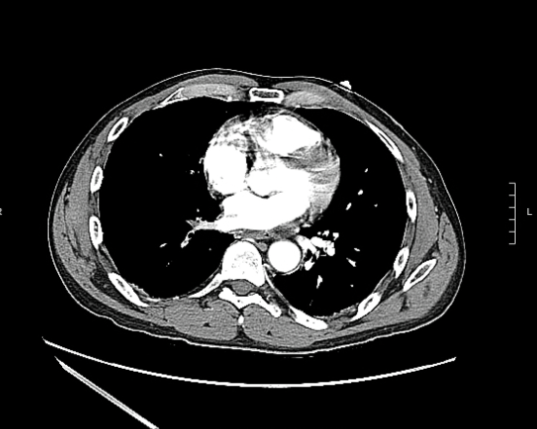
Case Presentation: A 48-year-old male with a past medical history of deep vein thrombosis (DVT) and pulmonary embolism (PE) presented with one day of severe chest pain, dizziness, and brief loss of consciousness resulting in a head injury. EKG showed T wave inversions in inferior leads and elevated troponin to 625 within a few hours. He was hemodynamically stable but had tachycardia. He was taken to the cardiac catheterization lab for a non-ST-elevation MI. Left heart catheterization which revealed no significant obstruction. CTA chest, which was done to rule out acute PE, revealed a thrombus within the posterior right pulmonary vein. The echocardiogram showed normal ejection fraction and normal right ventricle chamber size but mildly reduced function. He did not require any vascular or surgical intervention. On retrospective review of his medical records, he was hospitalized for a stroke 4 years prior to the presentation and was found to have multiple infarcts in right anterior cerebral artery and middle cerebral artery watershed areas and left cerebellar posterior inferior cerebellar artery territory on MRI brain. CTA head and neck revealed markedly stenotic right internal carotid artery and diminished right MCA flow. The transthoracic echocardiogram at that time did not show any intracardiac thrombus, vegetation or any right-to-left shunt. One year later, he was hospitalized once again for extensive left lower extremity DVT and a large right main and left subsegmental PE. Thrombophilia workup showed positive lupus anticoagulant but was negative for anticardiolipin, anti-beta2-glycoprotein antibodies, factor V Leiden, and prothrombin gene mutation. He was diagnosed with antiphospholipid syndrome (APS) which might have led to recurrent thromboembolism. The cryptogenic stroke at a young age with no apparent cardiovascular risk factors may have been precipitated by hypercoagulability. He was anticoagulated with heparin drip which was later bridged to warfarin on discharge. His chest pain resolved on outpatient follow-up.
Discussion: Pulmonary vein thrombosis (PVT) is extremely rare but can occur in the setting of hypercoagulable state such as APS, or in association with major lung surgery. Most of the literature is based on case reports. It presents itself with dyspnea, chest pain, hemoptysis, shortness of breath, and pulmonary congestion on X-ray. It can be difficult to diagnose clinically but is typically detected by CT angiogram, transesophageal echo, or MR venogram. It can result in life-threatening complications if the clot burden is significant, such as cardiogenic shock, cor pulmonale, pulmonary necrosis, and embolic stroke. Our patient’s stroke may have been caused by an embolism from PVT. However, there were no pulmonary symptoms or chest imaging at the time. There are no published treatment guidelines on optimal management, but systemic anticoagulation is the mainstay treatment. Vascular procedures such as thrombectomy might be required in severe cases. Antibiotic therapy may be indicated in cases with high clot burden due to risk of necrosis and secondary infection. Surgical resection of necrotic tissue is an option in patients with severe sepsis, or massive hemoptysis.
Conclusions: Clinicians should be aware that PVT is an underrecognized condition. It can present in a non-specific manner, and early diagnosis and treatment is crucial to prevent lethal complications. PVT should be considered a differential in patients with chest pain, especially in the setting of antiphospholipid syndrome.