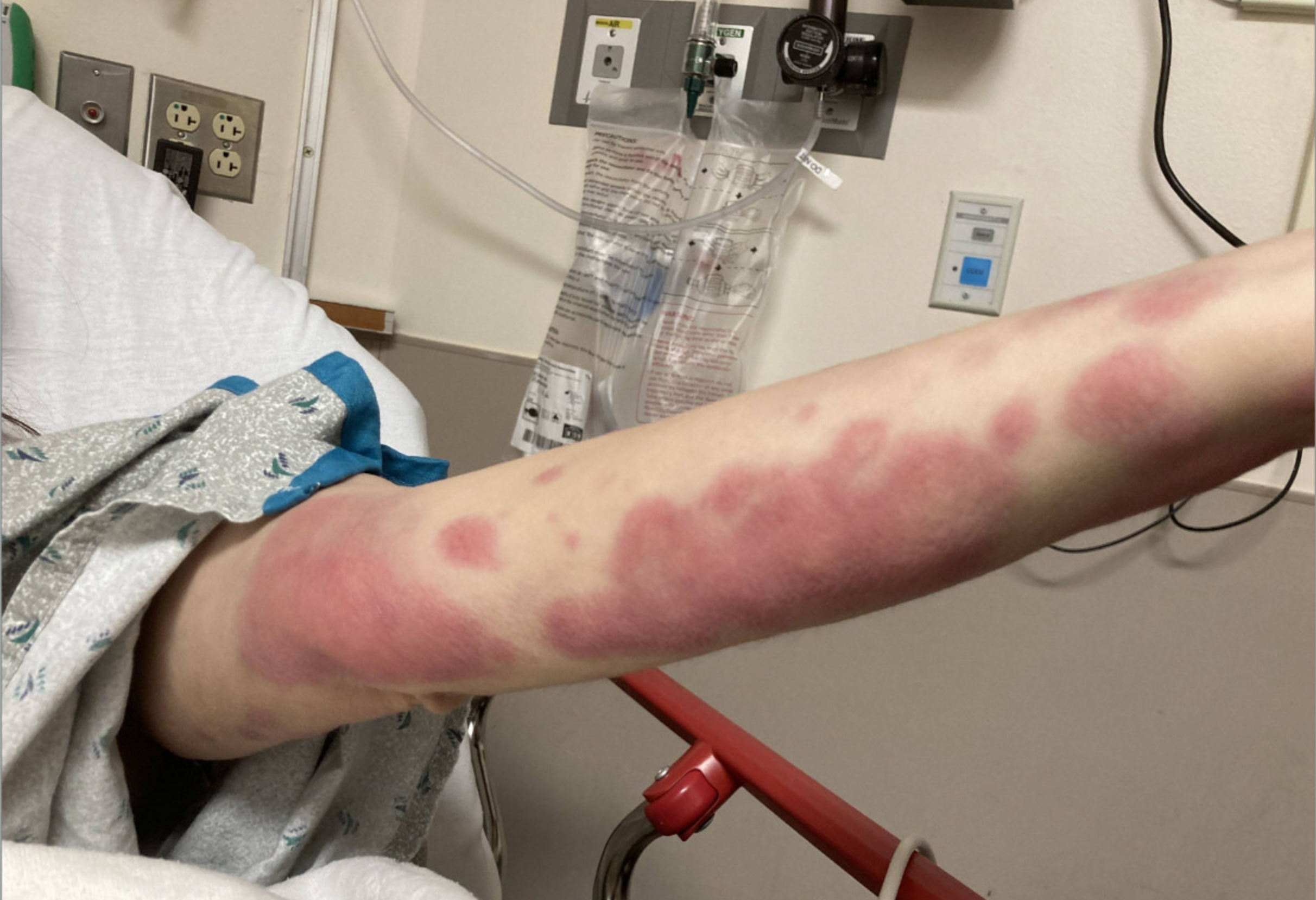
Case Presentation: A 38-year-old woman with no significant past medical history presented with fever, diffuse rash, and hypoesthesia. One year prior, the patient developed flesh-colored patches on her abdomen and raised lesions under her left arm. A biopsy revealed cutaneous nontuberculous mycobacterial infection with 4+ acid fast bacilli (AFB) stain, but AFB cultures were negative. She was treated with clarithromycin leading to symptom resolution. Months later, the rash returned as larger, mildly tender, erythematous, indurated circular lesions, mostly on her extremities. She then developed high fevers which prompted hospital admission. On exam, in addition to her skin lesions, she was noted to have loss of the lateral left eyebrow (madarosis). She reported patchy areas of hypoesthesia on her lower extremities. Admission diagnostics included negative viral panel, blood cultures, urinalysis, and normal chest x-ray. Skin biopsy revealed numerous atypical mycobacterial organisms and granulomatous dermatitis. The diagnosis of M. leprae was confirmed by PCR testing of the skin biopsy. Immune evaluation revealed CD3, CD4, CD8, and B-cell lymphopenia, low IgG, and absent lymphocyte proliferation to candida and tetanus. Whole-genome sequencing identified a novel GATA2 variant.She was started on treatment with monthly minocycline, rifampin, and moxifloxacin, alongside oral prednisone and methotrexate. She responded well with significant improvement in skin lesions and resolution of fevers, and had no worsening neuropathy.
Discussion: Hansen’s Disease (HD), caused by Mycobacterium leprae, is a rare disease characterized by a spectrum of dermatologic, neurologic, and systemic manifestations. Leprosy incidence has increased in the past decade in the United States, primarily in Florida (1). Global climate change may allow armadillos, the primary animal reservoir for M. leprae, to travel farther north. Leprosy is a diagnostic challenge in non-endemic regions due to its rarity and varied clinical presentations. Therapeutic regimens typically involve multi-drug therapy with dapsone, rifampin, and clofazimine over 6-24 months (2). Patients may also develop significant immune reactions to infection with M. leprae such as a reversal reaction with granuloma formation or erythema nodosum leposum—a vasculitis with immune complex deposits. Patients require treatment for these inflammatory processes with steroids, methotrexate, or thalidomide.GATA2 mutations are associated with susceptibility to opportunistic mycobacterial infections and increased risk for myelodysplastic syndrome (4). Our patient’s susceptibility may be attributed to her GATA2 heterozygous variant, potentially predisposing her to M. leprae. Immune evaluation revealed significant lymphopenia and hypogammaglobulinemia, aligning with the impact of the GATA2 variant on the patient’s immune function; however, further investigation is needed.
Conclusions: Our case illustrates the complex diagnosis and management of Hansen’s Disease in a non-endemic region, where global climate change threatens to increase incidence. The case highlights PCR testing as a key diagnostic tool, given the unreliability of traditional culture methods. Additionally, the presence of a GATA2 heterozygous variant emphasizes the need for further exploration of GATA2’s role in immunosuppression and susceptibility to mycobacterial disease.