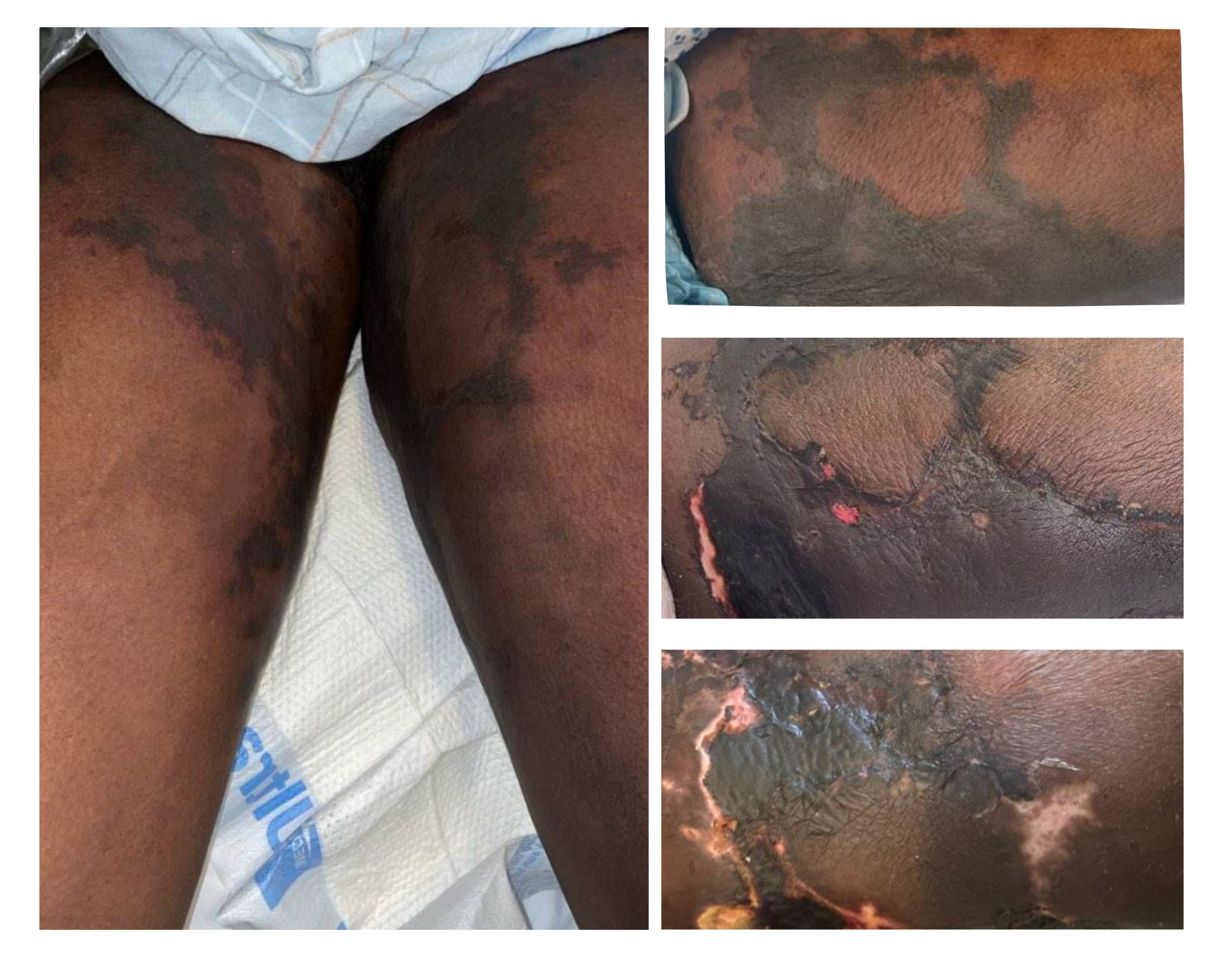
Case Presentation: A 50-year-old female with a history of CKD5, cervical cancer, and severe PAD presented to the ED with 1 day of altered mental status and hypoxia. On arrival, she was found to be in septic shock with renal failure. She underwent emergent hemodialysis (HD) and was admitted to the ICU. Imaging was consistent with pneumonia with cultures positive for Klebsiella and she improved rapidly after initiation of antibiotics. On hospital day three, she was transferred to the general medicine floor. Two days after transfer, the patient reported severe pain in her bilateral thighs and exam revealed a new symmetric retiform purpuric plaques/angulated purpura and patches. Dermatology and Vascular surgery were consulted. CT angiogram was done and showed no occlusion. Over the next few days, the rash progressed to include her bilateral breasts, as well as her right forearm. Biopsy results confirmed a diagnosis of calciphylaxis. The patient’s hospital course was further complicated by severe pain and difficulty tolerating HD. She was ultimately discharged to an inpatient hospice facility.
Discussion: This is a case about calciphylaxis and its unexpected presentation mid-hospital course. The initial differential diagnosis included calciphylaxis, primary occlusive vasculopathy, acquired coagulopathy, and septic vasculopathy. Calciphylaxis remained high on differential given the fatty area distribution and severe pain. Biopsy, tested using Von Kossa stain, demonstrated classic findings of calcium in small blood vessels in the subcutis and in the connective tissue in the deep dermis. The team’s early recognition of the rash’s appearance and characteristic distribution, as well as biopsy allowed for a timely diagnosis. Calciphylaxis is a rare, highly fatal condition usually seen in patients with ESRD. In our case, the patient had CKD 5 with acute worsening of her renal disease in the setting of sepsis requiring initiation of HD. The rash is characterized by cutaneous arterial calcification causing tissue ischemia, resulting in painful non-healing wounds. It usually affects areas with increased adipose tissue such as the thighs, buttocks, and abdomen, as was the case with our patient, whose rash began in the thighs and progressed to her breasts. Mortality usually results from ulcerated lesions leading to infection and sepsis; 1-year mortality rates are between 45-80%, with a mean survival time of 9.5 months. In the case of our patient, mortality occurred just under 2 months after diagnosis. Her cause of death was related to her discontinuation of HD. Unfortunately, many treatment modalities ranging from sodium-thiosulfate infusions to parathyroidectomies have been trialed in calciphylaxis without success. Early recognition of this disease allows patients to prioritize advanced care planning and for the clinical team to help ensure adequate pain control, which can be exceptionally difficult. Our patient required a hydromorphone PCA with both intermittent and continuous dosing, which was able to be continued when she was transferred to inpatient hospice.
Conclusions: Calciphylaxis should be considered in any patient with a rapidly progressive rash with primarily fatty area distribution, especially in patients with predisposing CKD history. Prompt referral to Dermatology is recommended so that biopsy can be obtained early. Finally, pain control and early goals of care discussion should be prioritized in patients with this incurable disease.