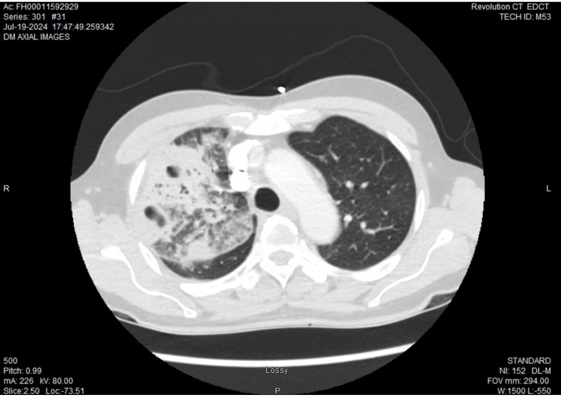
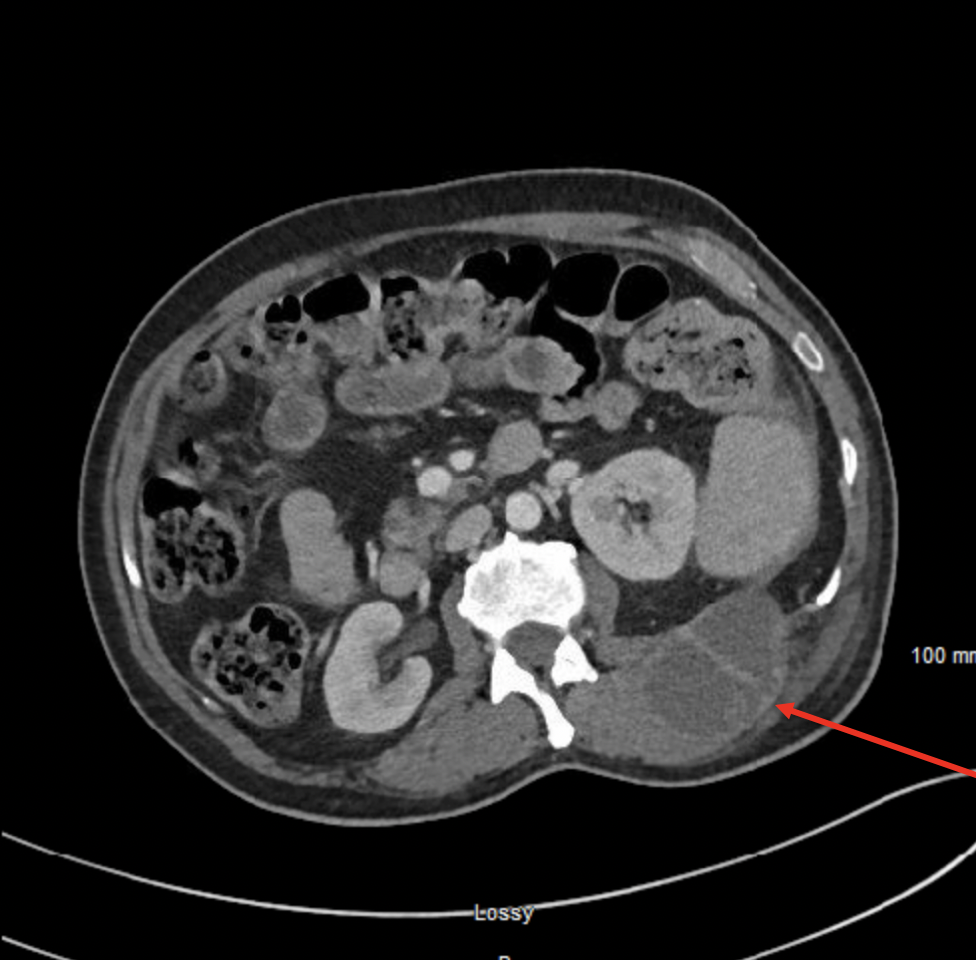
Case Presentation: The first case involved a 70-year-old Hmong-speaking man who presented with a few weeks of dry productive cough and worsening shortness of breath. He had decreased oral intake and postprandial fatigue with 20 pounds of unintentional weight loss in the preceding months. He was born in Laos but raised in Thailand, and had lived in Wisconsin for the past 20 years. Recent travel included a trip to Laos five months prior to admission. CT chest showed reticulonodular opacities in the right upper lobe of the lung with multiple cavitary lesions. Sputum cultures grew an isolate which was identified by MALDI-TOF-MS as Burkholderia thailandensis. Due to known cross-reactivity between B. thailandensis and Burkholderia pseudomallei, the isolate was forwarded to the Wisconsin State Laboratory of Hygiene for further testing, where it was identified as B. pseudomallei. After identification, antibiotics were transitioned to ceftazidime for 2 weeks, followed by a 90-day course of trimethoprim-sulfamethoxazole. Following treatment, the patient reported improvement and repeat chest X-rays demonstrated resolution of the right upper lobe cavitation. The second case involved a 58-year-old man who presented with worsening back pain for the preceding 2 weeks, accompanied by fever and anorexia. The patient had recently immigrated to the US from Nepal. His symptoms originally began a week prior to traveling. CT of the abdomen showed a multi-loculated mass originating in the abdominal wall, extending into the retroperitoneum, measuring 19 cm x 6 cm x 5.5 cm. Upon admission, the abscess was drained and yielded 350 mL of purulent fluid. Empiric piperacillin-tazobactam was initiated while awaiting culture results from the fluid. Given the suspicion of infection with B. pseudomallei, the antibiotics were switched to ceftazidime. Five days later, the laboratory workup confirmed infection with B. pseudomallei. Due to complicated social circumstances, he was not a candidate for the recommended 2 week ceftazidime therapy and instead was transitioned to a 90 day course of trimethoprim-sulfamethoxazole with overall improvement and resolution of the fluid collection on follow-up.
Discussion: B. pseudomallei is a bacterium that is endemic to southeast Asia and northern Australia but is rarely found in The United States. This pathogen results in melioidosis which usually causes asymptomatic carriage but can develop into severe infection with high mortality rates. Its varied presentation can be cavitary pneumonia, system wide abscesses, osteomyelitis, pneumonia, gastroenteritis, and/or meningitis. The manifestations can strongly mimic more common infections such as tuberculosis. Both patients likely acquired the bacteria by travel to endemic regions. However, within the last decade it has been found to be locally endemic to the Southeast US. CDC classifications label B. pseudomallei as a Tier 1 Category B Select Agent of Bioterrorism that requires strict lab handling and immediate eradication after identification. Current treatment guidelines require a two-phase treatment approach and prolonged follow-up to ensure disease eradication.
Conclusions: Although B. pseudomallei infection is often asymptomatic, the potential for melioidosis to become a life-threatening illness is underappreciated. Here we report two recent cases of melioidosis in the United States related to travel, highlighting its unpredictable presentation and the benefits of prompt detection and treatment.