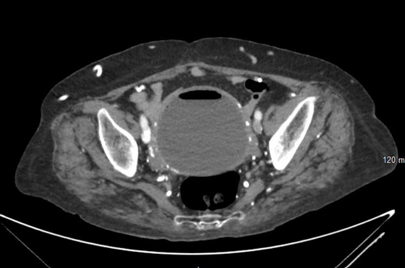
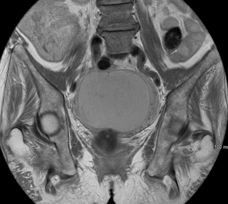
Case Presentation: A 57-year-old woman with resistant hypertension and end-stage renal disease on hemodialysis presented with fever, chills, and suprapubic pain of five days. Laboratory evaluation showed elevated wbc at 16 10*3/uL and hemoglobin 8.6 g/dL(at baseline). CT abdomen and pelvis showed a distended urinary bladder with mural thickening and antidependent air with additional foci extending along the bladder dome (Figure 1), raising suspicion for emphysematous cystitis. Blood cultures were collected and remained negative. Urine was unobtained as the patient had not made urine in many years and attempts at straight catheterization were unsuccessful. She was treated empirically with Zosyn. Given the appearance of imaging and the patient’s anuria, the diagnosis of emphysematous cystitis was questioned. Additional imaging with MRI showed a markedly distended endometrium with fluid and gas, concerning for endometritis vs. pyometra and an atrophic urinary bladder. Cultures collected from the purulent intrauterine fluid showed E. coli. She continued to have pain and lack of improvement despite undergoing appropriate antibiotic treatment and ultimately underwent exploratory laparotomy, hysterectomy, and bilateral salpingo-oophorectomy.
Discussion: Pyometra is a life-threatening infection resulting in the accumulation of pus in the uterus. This case involves a patient with acute onset of fever, chills, and suprapubic pain. Pyometra is often misdiagnosed due to its rarity and nonspecific symptoms. A diagnosis of emphysematous cystitis is more common and fit with the initial presenting symptoms, however it was questioned as it did not fit the patient’s clinical context. For hospitalists, it is crucial to personally review imaging and correlate findings to the clinical context of the patient they are caring for. This can help discern when further evaluation is necessary, especially when the diagnosis is incongruent with the patient’s history or exam. In this case, the imaging showed a distended bladder with urine, however the patient had been anuric for many years. Additionally, pyometra, although rare, should be included in the differential for postmenopausal patients presenting with nonspecific abdominal pain. Initial evaluation via ultrasound is preferred but imaging with CT or MRI is advised to confirm complications such as perforation ¹. Antibiotics may be a viable option for stable patients. However, given the high morbidity and mortality from perforation of pyometra, surgical management – including cervical dilation and drainage, endometrial aspiration, hysteroscopy, or total hysterectomy – should be considered emergently if the patient does not improve ²,³. Follow-up, including histological review, is advised as there is a possibility of cervical stenosis secondary to malignancy resulting in pyometra ².
Conclusions: Pyometra is a rare intrauterine infection that is often misdiagnosed in postmenopausal women due to its nonspecific presentation. Diagnosing and managing pyometra requires hospitalists to use appropriate imaging, interpret results in a clinically relevant context, and coordinate timely consultation with gynecology for management.