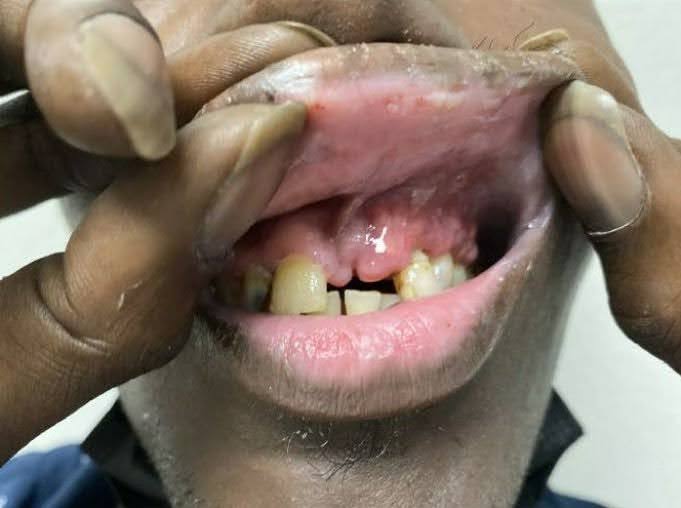
Case Presentation: The patient is a 37-year-old male with medical history significant for HIV infection. He was admitted for evaluation of a rapidly enlarging, left maxillary mass over the previous 6 months. At the time of presentation, it was estimated that he had not been on highly active antiretroviral therapy (HAART) for 12 months. His last known CD4 count was 43. Of note, he was evaluated at several outside hospitals for the same and told that he likely had Kaposi Sarcoma. He reported that biopsy was deferred due to pancytopenia, and he was recommended to continue his work-up in the outpatient setting. He was unfortunately lost to follow-up. On admission, initial labs were only significant for mild pancytopenia. A maxillofacial CT scan revealed a rounded soft tissue mass with bony erosion at the left maxilla with other radiologic findings consistent with Kaposi Sarcoma. Oncology and infectious disease consultations were requested at this time. Additionally, interventional radiology was consulted for tissue biopsy. Final pathology revealed an EBV+, ALK1- high-grade lymphoma with cMYC positivity. High grade FISH did not detect MYC reamplification or rearrangement; findings consistent with plasmablastic lymphoma. Given the aggressive nature of the tumor, the patient was transferred to an outside hospital to initiate chemotherapy. Under the direction of oncology, the patient was started on dose-adjusted EPOCH (etoposide, prednisolone, doxorubicin, vincristine, and cyclophosphamide). Additionally, HAART was initiated with bictegravir/emtricitabine/tenofovir AF as well as appropriate prophylaxis against opportunistic infections.Following 5 days of inpatient treatment with the above regimen, the patient was eventually discharged home for outpatient follow-up. He was seen in the outpatient clinic about one month following hospital discharge. Physical exam revealed notable improvement in the maxillary lesion. At this time, he was continued on DA-EPOCH given the positive response of his disease.
Discussion: Plasmablastic Lymphoma (PBL) is a rare and aggressive non-Hodgkin B-cell lymphoma often seen in immunocompromised population. Tissue biopsy is required for diagnosis in suspicious oral lesions to differentiate Kaposi Sarcoma and Plasmablastic Lymphoma. Unfortunately, delays in diagnosis of these oral lesions is well-documented due to multiple factors including previous misdiagnosis, stigmatized patients avoiding further work-up, and biopsy delay for various reasons. Once a diagnosis it achieved, treatment of this disease remains in its infancy. While various chemotherapeutic regimens have been used for the treatment of PBL since it was first described in 1997, the morbidity and mortality remains high. CHOP and DA-EPOCH remain the most commonly used regimens, but with advancements in the understanding of molecular targets, clinical trials are underway investigating alternate regimens.
Conclusions: It often falls onto the generalists within the hospital to advocate for patients to receive the medical attention that is needed. Recognizing and understanding the underlying social, society, and disease-state frameworks our patients deal with in the context of their illness allows hospitalists to better achieve that goal. Despite the involvement of several specialists, our hospitalist team was the backbone of treatment for this patient and allowed for smooth transition of care between the hospital and outpatient setting.