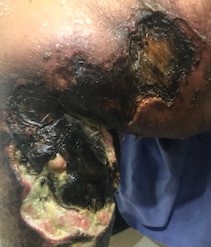
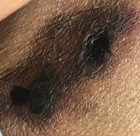
Case Presentation: A 47-year-old female with ESRD, chronic hypotension, anemia of chronic disease, depression and anxiety presented with painful chronic non-healing bilateral lower extremity wounds. Her wounds started about 3 months prior as superficial hyperpigmented painful areas which had gradually become indurated and subsequently ulcerated. Her wounds were extremely painful, limiting her ability to move and her overall functional status. She started to have other areas with hyperpigmentation in her bilateral thighs and breasts. She had elevated phosphorus and parathyroid hormone (PTH) which prompted transitioning from peritoneal dialysis (PD) to hemodialysis (HD). Her PTH of >1,000 pg/ml (12-88 pg/ml) was treated with Cinacalcet. On admission, her labs were consistent with ESRD and noticeably elevated Phosphorus of 7.8 mg/dl (2.5 – 5.0 mg/dl), Calcium of 10.5 mg/dl (8.6 10.3 mg/dl), PTH of 417 pg/ml and WBC of 13.6 (4 – 10 10E3/mcl). Her exam was unremarkable except two significant ulcerations to her legs. Her left leg wound measured 26 cm x 12 cm and her right leg wound measured 22 cm x 19 cm. Both wounds had poorly defined edges, scant brown and serosanguinous drainage and wound bed of mainly necrotic tissues. Both wounds also had thick adherent eschars and excruciatingly tender to palpation or touch (Figure 1). A punch biopsy showed fat necrosis and consistent with CUA.
Discussion: CUA develops as a result of occlusion of blood vessels in the subcutaneous fat and dermis. The lumen becomes narrowed by calcification in the media layer of the vessel walls and fibrosis underneath the intima and eventually thrombosis in the vessel lumen. This leads to ischemic cutaneous infarcts which can progress to severe ulcerations. Although it can manifest in non-dialysis patients, it is more common with patients who are on dialysis where hypercalcemia, hyperphosphatemia and hyperparathyroidism are common issues. Biopsy is not required but mainly is a clinical diagnosis with ESRD patients. Biopsy is likely needed to discern CUA from other skin lesions. Bone scan may be beneficial when biopsy is not feasible or inconclusive. Treatment for CUA is directed to stop calcification (correct hyperparathyroidism with Cinacalcet or surgery and aggressive HD to maintain good phosphorous/calcium level), promote decalcification (Na thiosulfate and vitamin K), wound care (debridement and hyperbaric oxygen therapy) and pain control. Due to the ischemic and neuropathic nature of the lesions, severe pain is a prominent feature of CUA. Often, pain is out of proportion to the skin lesions and can precede the appearance of skin lesions. Susceptibility to infection also makes CUA highly devastating with annual mortality of 40-80%.
Conclusions: The patient had the characteristic features and biopsy affirmative of CUA. Multidisciplinary approach was employed during her hospital course and she initially responded to the treatment modalities for CUA. However, she was transferred to a long-term care facility and subsequently developed complications from her wounds which led to her death. CUA is a life threatening and debilitating diagnosis to patients with ESRD, hence timely recognition is essential. Despite the low incidence of CUA, healthcare providers should be cognizant of its clinical features and management given the increase of ESRD patients.