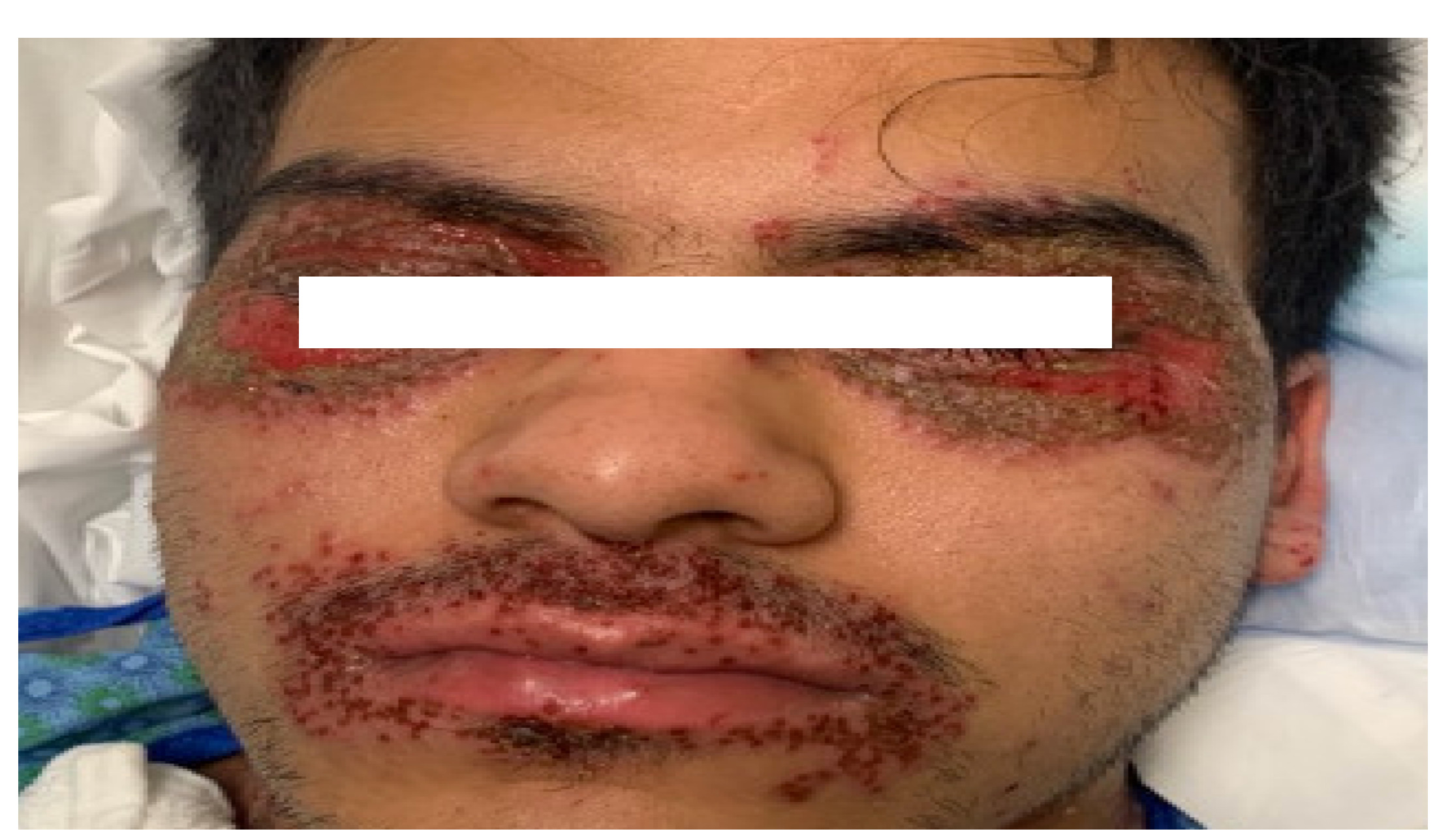
Case Presentation: A 22-year-old male with a history of atopic dermatitis presented with worsening of eczema lesions around his eyes and mouth with blurring of vision bilaterally despite being on oral antibiotics. 2 weeks ago he was treated with a steroid taper for eczema flare in his forearms. Patient reported that he has always had eczema patches around his eyes associated with puffiness and mild discomfort. 3 days prior to admission, he started to flare again around his mouth and eyes, with associated blisters and eye pain. He was seen in another facility where underwent CT of the orbits with contrast, notable for bilateral periorbital preseptal inflammatory changes without postseptal inflammation or discrete abscess formation. He was diagnosed with preseptal cellulitis and was started on mupirocin ointment, sulfacetamide ophthalmic solution and Keflex 500 mg BID x 10 days. Past history and family history are noncontributory. Patient is a non-smoker and drinks alcohol occasionally. On examination, he was febrile to 102 degrees, otherwise hemodynamically stable.Systemic examination was largely normal with exception to skin and eye examination.Skin examination revealed moderately thickened scaly lichenified plaques involving the antecubital and popliteal fossae bilaterally with scattered eroded papulesPeriorbitally and periorally, golden crusted plaques with studded eroded papules were present. Superficial eroded plaques were present infraorbitally and involving lateral canthi.Slit lamp examination of the eyes with fluorescein revealed several small, round corneal ulcers near the limbus consistent with staph marginal ulcers especially in the setting of blepharitis in both eyes His BMP and CMP were largely within normal limits. Patient was started on broad spectrum antibiotics and high dose IV acyclovir initially. Initial cultures sent from the eyes came back positive for Methicillin sensitive staph aureus. HSV 1 PCR also came back positive on Day 2. HIV test was negative. In addition to the above, topical antibiotics, steroid eye drops and hydrating creams for his eye lids were initiated. The IV regimen of antibiotics and antivirals and was changed to oral dose on Day 5 when his lesions had improved
Discussion: Patients with atopic dermatitis are at risk for developing an HSV-related skin complication called “eczema herpeticum”. It is also known as Kaposi’s varicelliform eruption. It is characterized by cutaneous pain and vesicular new skin lesions secondary to a viral infection (usually HSV-1). In most cases, the rash may be difficult to distinguish from the patient’s baseline eczema if it is poorly controlled, or can easily be dismissed as a bacterial superinfection. Eczema herpeticum can spread rapidly, leading to severe morbidity and mortality in the absence of antiviral therapy .Thus, prompt antiviral therapy is indicated while awaiting diagnostic testing for HSV, including administration of intravenous acyclovir in severe cases.
Conclusions: Atopic dermatitis is a common diagnosis in the outpatient setting. Impaired protective skin function, such as a flare facilitates HSV infection. Patients should be evaluated for secondary herpes virus infection due to complications associated with it. Secondary bacterial infection is also a common complication. Early awareness of eczema herpeticum and prescription of systemic antiviral medication are very important. Consultation with an ophthalmologist is also required for diagnosing herpes keratitis