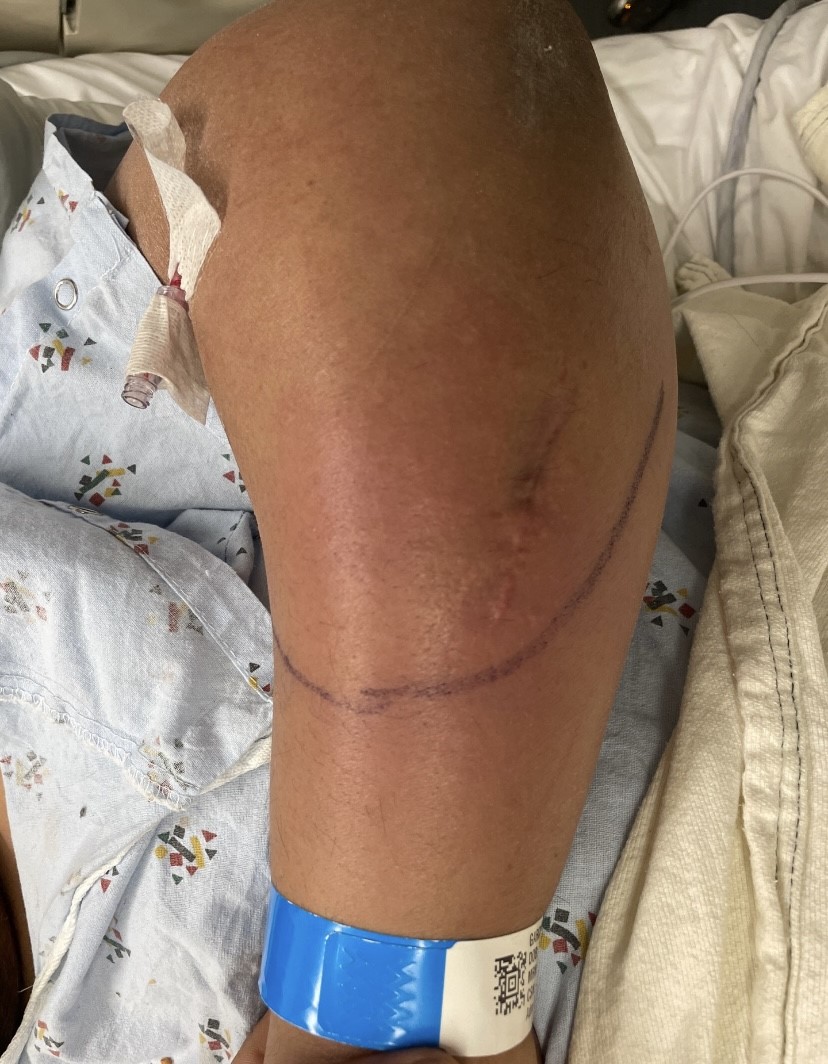
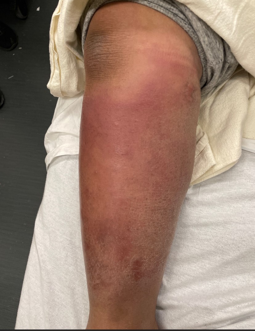
Case Presentation: A 40-year-old Hispanic male with a history of heart failure, hypothyroidism, and pre-diabetes was admitted for a warm, itchy, and painful rash on his extremities, accompanied by swelling in his upper limbs. He also experienced joint pain and stiffness in his elbows, knees, ankles, wrists, and back, without fever. Examination revealed pink, warm, indurated plaques with ill-defined margins on both forearms and legs, which were tender to touch. The rash was atypical for erythema nodosum, being pruritic, larger, and located beyond the shins. A skin biopsy was performed to differentiate between Lofgren syndrome, panniculitis, and cellulitis, confirming the diagnosis as erythema nodosum. Inflammatory markers, including CRP, ESR, and pro-calcitonin, were elevated, while the ACE level was normal. CT angiography revealed bilateral hilar and mediastinal lymphadenopathy, along with peri-bronchial consolidation in the left lower lobe. There was clinical concern for Lofgren syndrome presentation of sarcoidosis with erythema nodosum, inflammatory arthritis (including ankle involvement), and hilar adenopathy on chest CT. Infectious markers mimicking sarcoidosis, including tuberculosis, were ruled out. The patient had elevated histoplasma and chlamydia IgG antibodies. Patient did not respond to initial NSAID treatment and was eventually started on prednisone 20 mg daily. Follow-up in the rheumatology clinic two weeks post-discharge showed significant improvement in erythema nodosum and ankle pain, leading to a tapering and eventual cessation of steroids. Given the positive response to steroids and the clinical picture, the ID team had low concerns for histoplasma and chlamydia infections.
Discussion: Sarcoidosis is an inflammatory disease of complex nature which is characterized by formation of non-caseating granulomas in multiple organs in the body [1]. Though primarily affecting the lung, sarcoidosis has been noted to involve the skin, eyes, liver, muscle, heart, kidneys amongst other organs [1]. Clinically, sarcoidosis may present in two main forms-Lofgren’s syndrome and non-LS form. They are noted to have a distinct clinical course and features and have varied prognostic outcomes [1,2]. Lofgren syndrome is the form of sarcoidosis that may present more acutely [3]. It is characterized by the presence of erythema nodosum, hilar lymphadenopathy and bilateral ankle arthritis. Typically such patients are also febrile [3,4]. These symptoms may resolve spontaneously over time or be responsive to steroids and have a good prognosis. These features separate them from the non-LS form. The non-LS form usually has more advanced radiological stages at diagnosis, tends to be more progressive and may be slow to respond to even corticosteroid treatment [1,2].
Conclusions: While uncommon, a high clinical suspicion for Lofgren’s syndrome is crucial when patients present with the typical symptoms, potentially eliminating the need for a biopsy for a definitive diagnosis [5]. Erythema nodosum can present in atypical forms. Although the clinical triad is highly specific for acute sarcoidosis (95% specific [6]), it’s essential to rule out conditions that mimic it, including tuberculosis, lymphoma, streptococcal infection, histoplasmosis, coccidiomycosis, mycoplasma, and Chlamydia pneumoniae infections [6]. Most cases are self-limited. However, in those patients who have persistent symptoms, a trial of either NSAIDS or steroids may contribute to quick recovery for the patient.