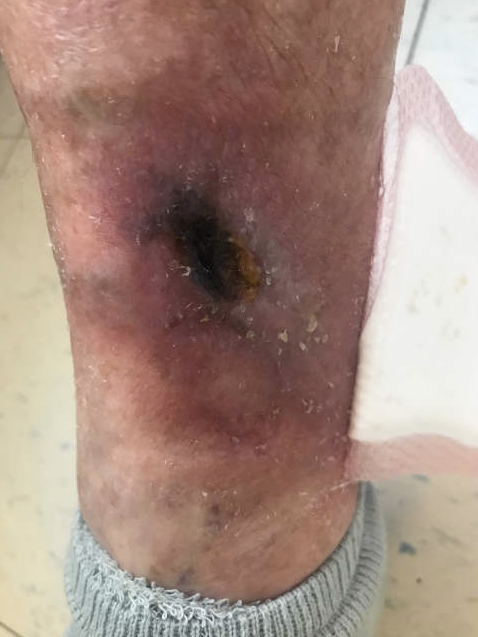
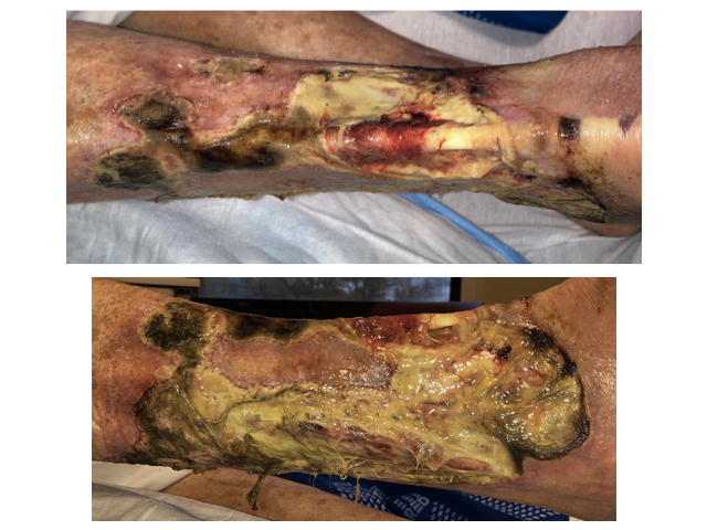
Case Presentation: 87-year-old man with a past medical history of wild type ATTR cardiac amyloidosis and atrial fibrillation, presented with a progressive right lower extremity ulcer. The wound initially started as a “pinhole” after dropping a toothpaste cap on the calf and progressed in the next three months despite multiple courses of antibiotics and wound care. A wedge skin biopsy obtained revealed stasis dermatitis with ulceration, acute inflammation, focal tissue necrosis and negative Congo Red staining. He was empirically treated for pyoderma gangrenosum with prednisone for 17 days, without clinical improvement. On admission, the wound had progressed to a large ulcer extending from lateral mid-shin to right ankle with significant peripheral necrosis and green malodorous discharge, exposing the distal half of the anterior tibialis tendon. Notable laboratory findings included a white blood cell count of 21×10^9/L and CRP of 77mg/L. A repeated punch skin biopsy revealed deep dermal and subcutaneous tissue calcification and intimal proliferation suggestive of calciphylaxis. Wound cultures grew pseudomonas aeruginosa and proteus mirabilis. Creatinine, liver function, calcium, parathyroid hormone, vitamin D-25, Protein C and S levels, ANA, cryoglobulin, ANCA, ankle brachial index and CT pan scan were all unrevealing for secondary causes. The only identifiable risk factor was systemic corticosteroid use, but its initiation was after the first wound appearance. The patient was started on sodium thiosulfate infusions, and was covered with cefepime for possible overlying cellulitis. The wound was debrided with subsequent autogenous skin graft implant.
Discussion: Chronic leg ulcers are most commonly caused by venous stasis. Microvascular disease such as NUC is a rare cause and the sensitivity of diagnosis on the first biopsy is only 50.5% [1]. Pyoderma Gangrenosum as a diagnosis of exclusion is also known to mimic multiple diseases [2] but typically responds to immunosuppressants within a month [3]. Calciphylaxis is most often seen in patients with end-stage renal disease on dialysis, but when it occurs in patients without advanced kidney disease it is known as Non-Uremic Calicphylaxis (NUC). The exact pathogenesis is unclear but is associated with cutaneous arteriole calcification and thrombotic occlusion. There is no unifying trigger in NUC, however female gender, underlying malignancy, chronic liver disease, prothrombotic conditions, autoimmune or granulomatous diseases, warfarin or corticosteroid use, as well as parathyroid hormone or Vitamin D abnormalities are recognized risk factors. For treatment, other than avoiding exacerbating factors such as systemic steroids, sodium thiosulfate, which increases the solubility of the vascular calcium deposits, is also frequently used off label. NUC is associated with a high one year mortality rate (up to 52%) most frequently due to complications from sepsis [4]. This patient was discharged on sodium thiosulfate treatment with ongoing wound care and a guarded prognosis.
Conclusions: While non-uremic calciphylaxis is a rare and poorly understood cause of skin necrosis, this case demonstrates the importance of hospitalists expanding and vigilantly reviewing the differential diagnosis when treatment response is not as expected, despite lacking other risk factors.