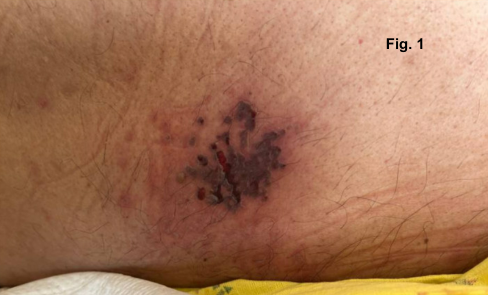
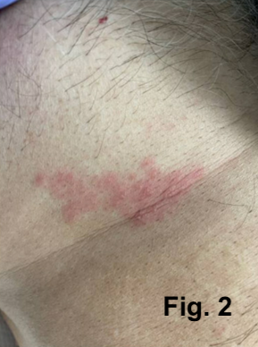
Case Presentation: A 62-year-old man with a history of diabetes presented to the emergency department with altered mental status (AMS) and increased urinary frequency. The patient was conscious but oriented only to self. Initial evaluation revealed hyponatremia (serum sodium: 125 mmol/L) with mild hyperglycemia (186 mg/dL). Further assessment of hyponatremia showed a serum osmolality of 263 mosm/kg, urine sodium of 111 mmol/L, and urine osmolality of 838 mosm/kg. A CT scan of his head showed no acute abnormalities, although parenchymal volume loss was noted. The patient was treated with hypertonic saline, oral urea, a loop diuretic, and fluid restriction (800 mL/day) for presumed syndrome of inappropriate antidiuretic hormone secretion (SIADH) based on urine osmolality findings. Early in his hospitalization, the patient developed scalloped, grouped erosions on his back (Fig. 1) and coalescing vesicles on an erythematous base across his right flank (Fig. 2). A PCR assay for varicella-zoster virus (VZV) on a skin swab returned positive, prompting initiation of intravenous (IV) acyclovir at 10 mg/kg every 8 hours. Blood serology revealed elevated VZV-specific IgG with normal IgM, consistent with viral reactivation. MRI of the brain showed atrophy without acute findings. Lumbar puncture revealed an opening pressure of 15 cm H2O, with cerebrospinal fluid (CSF) analysis showing elevated protein (79 mg/dL), increased nucleated cells (205/μL with 90% lymphocytes), and elevated glucose (144 mg/dL). CSF PCR assay for VZV was positive, leading to a diagnosis of disseminated VZV with encephalitis. IV acyclovir was continued. The patient’s clinical condition improved, serum sodium levels normalized, and a repeat lumbar puncture after 14 days of treatment was negative for VZV on CSF PCR. IV acyclovir was discontinued, and the patient was discharged in stable condition with mental status restored to baseline.
Discussion: While typically relatively benign, VZV infections can lead to significant morbidity in certain patient populations. Advanced age and diabetes are established risk factors for VZV reactivation. Among the potential complications is the development of SIADH, which can result in severe hyponatremia. The coexistence of VZV infection and SIADH has been documented in the medical literature, although it remains a relatively rare complication, with only a limited number of cases reported. The exact mechanism by which VZV induces SIADH is not yet fully understood. A prevailing hypothesis suggests that VZV, which resides latently in the sensory neurons of the dorsal root ganglia, may spread to peripheral osmoreceptors during reactivation. These osmoreceptors, which transmit signals via the dorsal root ganglia, could be disrupted by the virus, thereby interfering with antidiuretic hormone (ADH) secretion. In our patient, the concurrent resolution of hyponatremia and disseminated VZV, along with the absence of other identifiable causes of SIADH, supports a potential association between SIADH and VZV. Notably, the patient’s AMS also improved with the correction of hyponatremia.
Conclusions: This case underscores the importance of clinicians maintaining a high index of suspicion for SIADH in patients with VZV infections who present with hyponatremia. Close monitoring and careful management of electrolyte imbalances in these individuals are essential to prevent potentially severe neurologic complications.