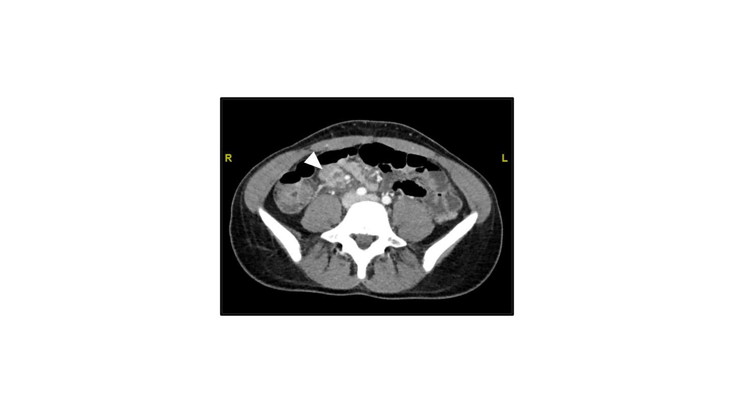
Case Presentation: A previously healthy 20-year-old presented with 5 days of fevers and diffuse abdominal pain. Her exam was notable for tachycardia to 118/min, fever of 103F with marked abdominal tenderness. Her laboratory results were significant for leukopenia (3.7 K/uL), anemia (9.2 g/dL), elevated D-dimer to 2.98 ug/ml, CRP of 56.33 mg/L, and troponin of 1.142 ng/ml. Computed tomography of the abdomen/pelvis demonstrated several necrotic small bowel mesenteric lymphadenopathy 2.7 x 2.1 cm with gallbladder wall edema. She was subsequently started on intravenous antibiotics. During her hospitalization, she continued to have daily cyclical fevers with persistent abdominal pain complicated by bilateral pleural effusions and meningeal symptoms. An extensive laboratory workup was unremarkable except for Epstein-Barr virus capsid antigen IgG, Parvovirus IgG, Human herpes virus-6 IgG antibody positivity. As the laboratory findings were unrevealing, she subsequently underwent a laparoscopic lymph node biopsy – that revealed severe necrotic lymphadenitis. During the rest of her hospitalization, the patient improved on conservative management. She was discharged home on day 13 in stable condition. Her final diagnosis was Kikuchi’s disease with an atypical location of lymphadenitis.
Discussion: Kikuchi disease is a rare, benign condition of unknown etiology usually characterized by cervical lymphadenopathy and fever. Rarely lymphadenopathy may be localized to a single lymph node or sometimes only mediastinal or retroperitoneal nodes may be involved [1] [2]. The most common clinical presentation of Kikuchi disease is fever and cervical lymphadenopathy. Fever is the primary symptom [3]. Since it is a diagnosis of exclusion the diagnosis is unlikely to be considered before biopsy in such patients who may present with a fever. No effective treatment has been established for Kikuchi disease [4]. Signs and symptoms usually resolve within one to four months [4]. Although it is self-limiting, recurrences have been reported in multiple cases [5,6,7,8]. Our case displays a rare case of Kikuchi with an atypical location of the lymph node. Fever being the primary symptom was present throughout the course. The location of the necrotic mesenteric lymph node near the small bowel is quite rare. As seen here the atypical presentation including transiently elevated troponins, pancytopenia [9,10,11], bilateral pleural effusions [12] and meningitis-like symptoms [13,14,15] can be seen. Patients should be screened for autoimmune conditions like systemic lupus erythematosus [3,16] and recurrences that can develop later in the disease [10].
Conclusions: Kikuchi disease, while rare, is important to consider in the differential diagnosis of a patient with fever and lymphadenitis. This case demonstrates an atypical presentation of the disease due to the anatomical location of lymphadenopathy. It’s important to recognize it early to prevent morbidity associated with the disease.