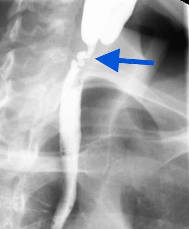
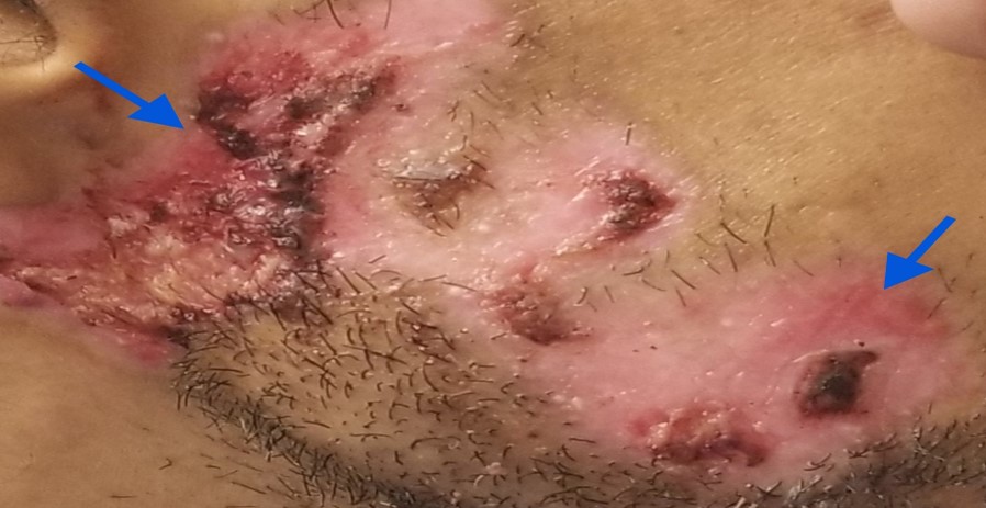
Case Presentation: A 37-year-old male with a history of bullous pemphigoid, sickle cell trait, and smoking, presented with 5 months of progressive dysphagia, oral lesions, and weight loss. Initiation of swallowing was intact, however the patient felt as though food was getting stuck mid- esophagus. The dysphagia began with solid food and progressed to liquids.Physical exam revealed a thin male with several active bullous pemphigoid lesions on the face and chest and poor oral hygiene, along with further lesions on the hard palate, gingiva, and posterior oropharynx. An esophagram revealed a severe serpiginous stricture in the cervical esophagus (C4-5) approximately 3 cm long.An EGD with an XP/ stricture endoscope showed multiple ulcerations and bullae in the upper esophagus; however the stricture was too narrow for the endoscope to pass the hypopharynx. Despite a course of IV steroids, repeat EGD was unsuccessful.Esophageal dilatation was considered, however the patient was deemed too high risk, so a gastroscopy tube was placed.
Discussion: Bullous pemphigoid and mucous membrane pemphigoid are rare autoimmune conditions typically seen in the elderly.Both conditions can affect the skin and mucosa as immunoglobulins and complement proteins are deposited in the epidermal/ mucosal basement membrane, promoting subepithelial blister formation. 85% of patients with mucous membrane pemphigoid have lesions in the oral cavity, but only 4% have esophageal involvement.Treatment consists of glucocorticoids and occasionally biologics or immunosuppressants.Esophageal involvement may require an enteral feeding tube when esophageal dilation in not an option.
Conclusions: Bullous pemphigoid and mucous membrane phemphigoid are rare conditions.The above case highlights an atypical case of both bullous pemphigoid and mucous membrane pemphigoid occurring simultaneously in a young patient.