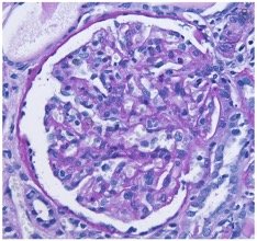
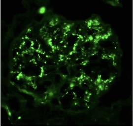
Case Presentation: A 28-year-old male with hypertension, hyperlipidemia, obstructive sleep apnea, and obesity presented with epistaxis, initially attributed to poorly controlled blood pressure. Laboratory evaluation revealed acute kidney injury and nephrotic-range proteinuria. He reported progressive dyspnea, lower extremity edema, and intermittent hemoptysis beginning two months earlier following a viral respiratory infection. Given his clinical course, low complement levels, and recent infection, the initial diagnosis was post-infectious glomerulonephritis (PIGN).Initial labs showed serum creatinine 1.71 mg/dL, albumin 2.1 g/dL, proteinuria 14 g/day, microscopic hematuria with RBC casts, and markedly reduced C3 (31 mg/dL) with normal C4. ANA, ANCA, and serum protein electrophoresis were negative. Renal ultrasound revealed mild left intrarenal collecting system dilation without hydronephrosis.Renal biopsy demonstrated severe mesangial and endocapillary hypercellularity, mild interstitial fibrosis and tubular atrophy (10–20%), and strong C3 deposition (3+) with minimal IgG (1+), kappa (1+), and lambda (trace). Electron microscopy revealed extensive mesangial electron-dense deposits, rare subendothelial deposits, and early membranoproliferative changes consistent with C3 glomerulopathy (C3GN).The patient received intravenous methylprednisolone (250 mg daily ×3 days), followed by oral prednisone (60 mg daily) and mycophenolate mofetil (1 g twice daily). Supportive therapy included diuresis, gastrointestinal prophylaxis, and blood pressure management. Over months, complement normalized and proteinuria improved from 14 g/day to 0.91 g/day. Immunosuppressive therapy was tapered, and complement inhibition with Iptacopan (Fabhalta) was initiated, achieving sustained clinical improvement.
Discussion: This case highlights diagnostic overlap between PIGN and C3GN, especially after infection with hypocomplementemia. Both share hematuria, proteinuria, and low C3 with normal C4, along with similar histology. Distinguishing features favoring C3GN include persistence or recurrence of proteinuria despite normalized complement, predominant C3 deposition with minimal immunoglobulin, and mesangial/subendothelial electron-dense deposits with early membranoproliferative changes.Prior viral infection likely triggered dysregulated alternative complement pathway activation. Management remains challenging due to variable pathogenesis and limited evidence-based therapies. Corticosteroids and mycophenolate are commonly used; emerging complement inhibitors such as Iptacopan show promise for refractory disease.
Conclusions: Renal biopsy is essential in patients presumed to have PIGN who exhibit relapsing proteinuria or persistent hypocomplementemia. Accurate differentiation of C3GN enables timely immunomodulatory and complement-targeted therapy, preventing progression to end-stage kidney disease and improving long-term outcomes.