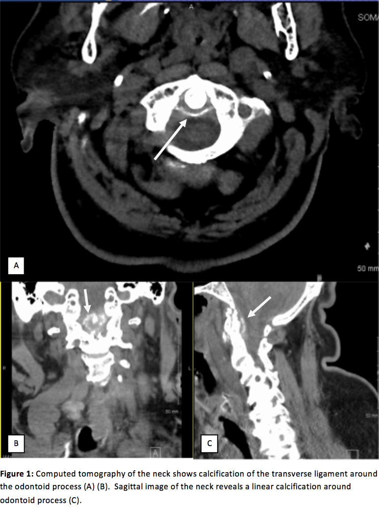
Case Presentation: A 81 year old Asian woman with past medical history of diabetes, hypertension, coronary artery disease, and arthritis presented with fever and five days of worsening neck stiffness and pain, worsened by neck movement. Laboratory examination revealed mild leukocytosis, CRP 28 and ESR 114. Broad spectrum antibiotics were started for possible meningitis. Lumbar Puncture, Encephalitis studies, ANCA, ANA and Rheumatoid factor were negative. Ferritin, Calcium and Uric Acid were within normal limits. Head and neck imaging did not reveal abscesses or signs of compression. High dose IV steroids were started two days after admission due to concern over anaphylaxis from antibiotics, with marked improvement in neck pain and range of motion after three doses. She was then transitioned to oral glucocorticoids and was discharged. Review of initial CT scan on admission illustrated crown-like density around the odontoid process.
Discussion: Crowned Dens Syndrome (CDS) is a disease characterized by acute cervical pain, fever, neck stiffness, elevated inflammatory markers, and radiologic calcifications of ligaments around the odontoid process. The syndrome was first described by Bouvet, et al in 1985 in four female patients with acute upper neck pain and imaging showing radio-opaque densities surrounding the odontoid process like a crown or halo. The authors hypothesized the deposits were made of both hydroxyapatite and calcium pyrophosphate dehydrate (CPPD) crystals, based on radiologic data correlating patients with known CPPD deposition disease to calcifications around the odontoid process. The diagnostic gold standard to identify peri-odontoid calcifications is CT tomography, while MRI is indicated for neurologic complications. CPPD deposition typically occurs in fibro and hyaline cartilage, and can cause mono or oligroarthritis in large joints such as the knee, wrist, shoulder and hip. Patients with known chondrocalcinosis may have asymptomatic calcification of the periodontoid region, but deposits in cruciform, transverse, alar and apical ligaments typically give rise to the symptoms of CDS. Rarely, CDDP deposition at the ligamentum flavum can cause compressive cervical myelopathy. Patient demographics with CDS is skewed toward women greater than seventy years old.
Standard treatment of CDS is NSAIDS or colchicine, with symptom resolution in days to weeks. Glucocorticoids may be used in severe cases and were successful in our patient. Combined therapy with prednisolone and NSAIDs may be the most effective treatment. Successful decompression surgery on elderly patients with CPPD deposition at the C1-C2 level causing functional quadriplegia, and another patient with cervical myelopathy, indicates the need for careful neurologic monitoring of CDS patients and low threshold for surgical evaluation. Radiologic follow-up with CT scan has shown decrease in size and even disappearance of calcifications within months to years.
Conclusions: CDS is a rare disease that can be misdiagnosed as meningitis, epidural abscess, rheumatoid arthritis, metastatic spinal tumor, polymalgia rheumatic and giant cell arteritis. Although painful neck on rotation has been proposed as a diagnostic tool to differentiate between CDS and meningeal irritation, patients with CDS undergo extensive testing as well as potentially harmful procedures. Our case illustrates a high clinical suspicion for CDS must be maintained, especially in elderly women, when workup of other etiologies is unremarkable.