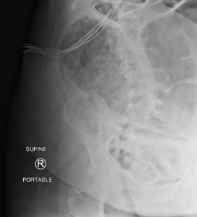
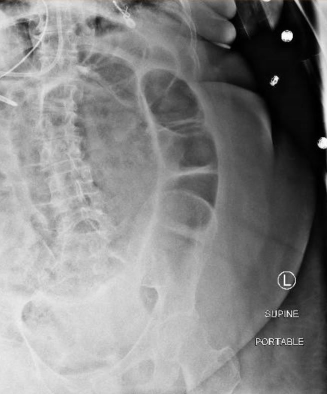
Case Presentation: A 70 year old female with past medical history of COPD on 2 liters oxygen at baseline presented with acute hypoxic respiratory failure due to COPD exacerbation secondary to metapneumovirus infection. Her hospitalization was prolonged from Methicillin sensitive Staphylococcus aureus pneumonia which required prolonged intubation, antibiotics and finally a tracheostomy. She was noted to be encephalopathic secondary to hypoxia and extremely constipated with no bowel movement in 16 days despite being on Colace twice daily. 3 weeks into hospitalization, her lower extremities were noted to be mottled with preserved peripheral pulses. Her abdomen was found to be distended and rigid, however tenderness could not be assessed as she remained encephalopathic despite being off sedation for days. Her urine output started declining and her peak pressures were intermittently elevated to 40 cm H2O on the ventilator. She became acutely hypotensive and her peripheral pulses were no longer felt. Her intra-abdominal pressure was measured through the Foley catheter and was found to be 39 cm H2O. Emergent surgical decompression performed at bedside for acute abdominal compartment syndrome. She was found to have bowel ischemia requiring resection and anastomosis.
Discussion: Abdominal Compartment Syndrome (ACS) is a rare complication in the medical setting. Risk factors include burns, trauma, massive ascites, intraperitoneal bleeding, pancreatitis and other medical illness requiring extensive fluid resuscitation. It is under-recognized because it primarily affects patients who are already critically ill with multi-organ dysfunction. More so, it is clinically silent until irreversible end organ damage has already occurred.Recognizing intra-abdominal hypertension (IAH) early is important so it can be treated before progressing to ACS. On physical exam, patients with ACS have a tensely distended abdomen but despite this, abdominal examination is a poor predictor of ACS. Other indicators of ACS include progressive oliguria, increased ventilator requirements, elevated central venous pressure, hypotension, tachycardia, jugular venous distention, peripheral edema, and acute pulmonary edema. Measurement of intra-abdominal pressure using a bladder catheter is the preferred method of diagnosis and it is essential that physicians are aware of this simple bedside test. In this case, our patient’s gut ischemia was attributed to new atrial fibrillation developed earlier during her admission and was possibly worsened by her severe constipation.
Conclusions: A high degree of suspicion and knowledge regarding diagnosis is required to provide specific treatment and prevent organ dysfunction in abdominal compartment syndrome.