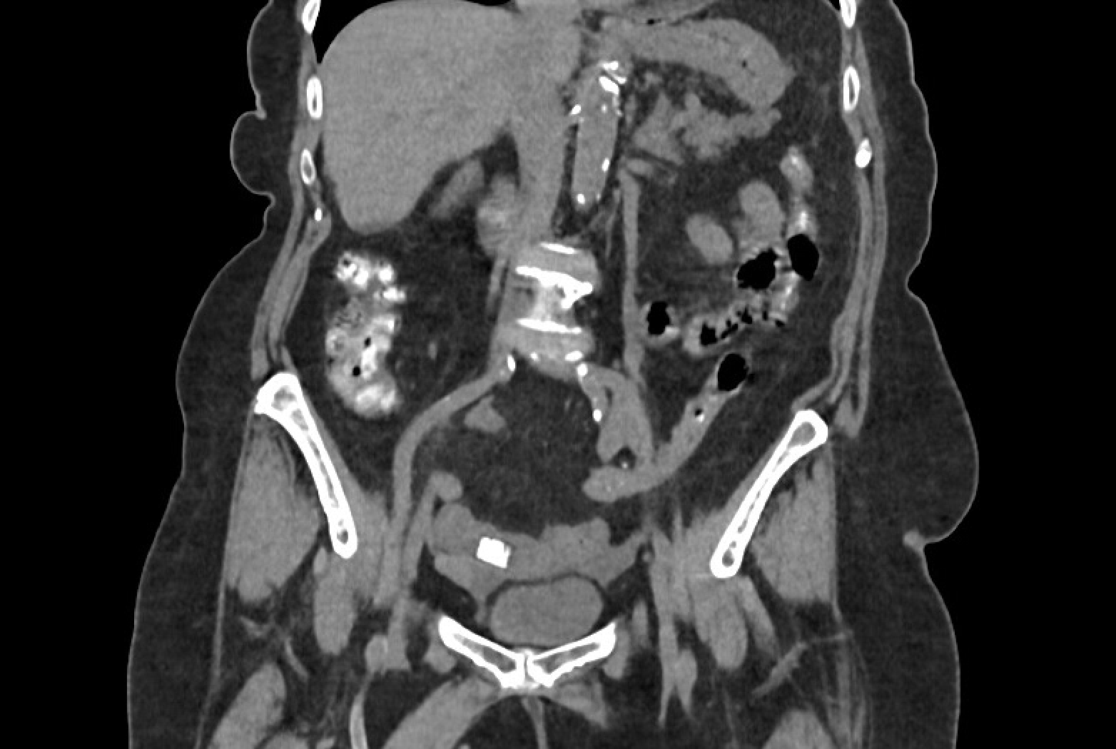
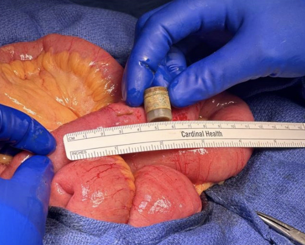
Case Presentation: An 85-year-old woman with a history of heart failure, stroke, persistent atrial fibrillation, and recent myocardial infarction treated with a drug eluting stent presented to urgent care with 4 days of abdominal pain, loss of appetite, vomiting, and PO intolerance. Exam revealed tenderness in the right upper quadrant but no guarding or rebound tenderness. CT scan showed a small bowel obstruction with a small radio-opaque intra-luminal mass (Figure 1). She was diagnosed with a small bowel obstruction with pill bezoar as the suspected etiology due to her taking 25 pills daily, including 14 supplements which she recently increased following her myocardial infarction. She initially improved with conservative management but failed advancement of her diet. A repeat CT scan showed dilation of the small bowel extending to the level of a radio-dense intraluminal object and potential small bowel pneumatosis proximal to the obstruction. The patient underwent urgent exploratory laparotomy with removal of a foreign body which was identified as a small cylinder of desiccant commonly found in pill bottles (Figure 2). Her post operative course was complicated by continued difficulty maintaining adequate oral intake requiring both parenteral nutrition and enteral tube feeds. She was eventually discharged with close follow up and instructions to stop multiple supplements.
Discussion: Pill bezoars are extremely rare and should be suspected when risk factors such as dysmotility, severe coexisting illness, and the use of opioid medications are present (1, 2). Excessive use of vitamins and natural products has been implicated in the formation of pill bezoars, but primarily in the setting of impaired GI motility (2). This case illustrates the need to keep foreign body ingestion on the differential in the elderly. Accidental ingestions show a bimodal age distribution with one peak in children and a second peak in adults over 80 years old (3). Fish and chicken bones are the most common foreign bodies in the elderly, followed by dental prosthesis, and medicine packaging. Risk factors include impaired swallowing, impaired intraoral sensation, mis-swallowing due to poor vision, and denture use (4). Most foreign body impactions occur in the cervical esophagus, presenting with symptoms of choking, coughing, drooling or sudden refusal of oral intake (4). If esophageal foreign body is suspected, upper endoscopy allows for prompt diagnosis and removal. Foreign bodies in the lower GI tract are more likely to pass on their own but may present with small bowel obstruction as in our patient. CT scans are a useful diagnostic tool for localizing and characterizing foreign bodies. It’s imperative to maintain a high clinical suspicion for foreign body ingestions in the right setting, as the risk for complications increases with delays in removal (4).
Conclusions: It’s important for hospitalists to be aware of the risk factors and possible clinical presentations of foreign body ingestions in the elderly as delays in diagnosis and foreign body removal significantly increase the risk of complications.