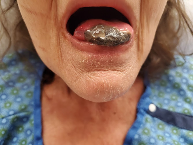
Case Presentation: A 60-year-old female with a history of tobacco use disorder, laryngeal cancer status post chemotherapy and radiation, hyperlipidemia, and hypertension presented with tongue pain. Ten days before her presentation, she developed a small painful blister on her tongue, which was associated with difficulty eating and drinking. She decided to come to the hospital when her blister started to change color, and the tip of her tongue became black. She denied associated fevers, difficulty breathing, vision changes, headaches, weakness, jaw pain, or rashes. In the ED, her vitals were significant for mild tachycardia (110 bpm). Her labs were significant for leukocytosis (13.5 K/cmm), hyponatremia (126 mEq/L), and elevated CRP (13.8 mg/L) with normal ESR (25 mm/h). Blood cultures were obtained, and the patient was started on empiric vancomycin, piperacillin-tazobactam, and clindamycin. A contrast-enhanced CT showed no enhancement at the tip of the tongue and no internal locules of gas. Given concern for possible thromboembolic disease, she was started on heparin, in addition to aspirin and atorvastatin. Otolaryngology performed a flexible laryngoscopy, which was unremarkable. Punch biopsy of the lesion showed an ulcer and granulation tissue with numerous bacterial microorganisms. Her antibiotics were discontinued on hospital day 4, as her infectious workup including blood cultures remained unremarkable. CTA of the head and neck was obtained which showed multifocal noncalcified plaque of left common and left internal carotid artery. Rheumatology work-up including C-ANCA and P-ANCA was negative, and a temporal artery biopsy did not show any evidence of temporal arteritis. The patient’s lesion remained stable throughout her hospitalization. She was discharged home with the diagnosis of lingual necrosis secondary to embolic atherosclerosis.
Discussion: Tongue necrosis is a rare clinical diagnosis and may be caused by a wide array of pathologies, including systemic vasculitis (giant cell arteritis, ANCA-associated vasculitis), malignancy (sarcoma or lymphoma), cardiovascular entities (hemorrhage, embolism, and cardiac arrest), infection (TB and syphilis), and medication use (vasopressin and ergotamine).(1) The primary vascular supply to the tongue is the lingual branch of the external carotid artery. There is also contralateral flow via the submucosal plexus and flow from the ipsilateral internal carotid artery.2 The rich vascular supply makes tongue infarction due to vascular phenomena a rare occurrence.(2) Our patient with a history of local radiation due to head and neck cancer, and atherosclerotic risk factors including smoking, hypertension, and hyperlipidemia, demonstrates a unique example of embolic necrosis of the tongue. Clinicians should maintain a high degree of suspicion for an embolic cause of necrosis in patients with multiple risk factors for atherosclerosis, especially in patients with a history of prior radiation. Our patient was treated with heparin and was discharged home with aspirin and a statin to reduce her thromboembolic disease risk.
Conclusions: Given the varied potential causes of tongue necrosis, we emphasize the importance of a cross-disciplinary team-based approach to patients who present with lingual necrosis. Our patient’s case was self-limited but, in cases where the underlying diagnosis is related to vasculitis, prompt treatment can prevent serious and irreversible end-organ damage.