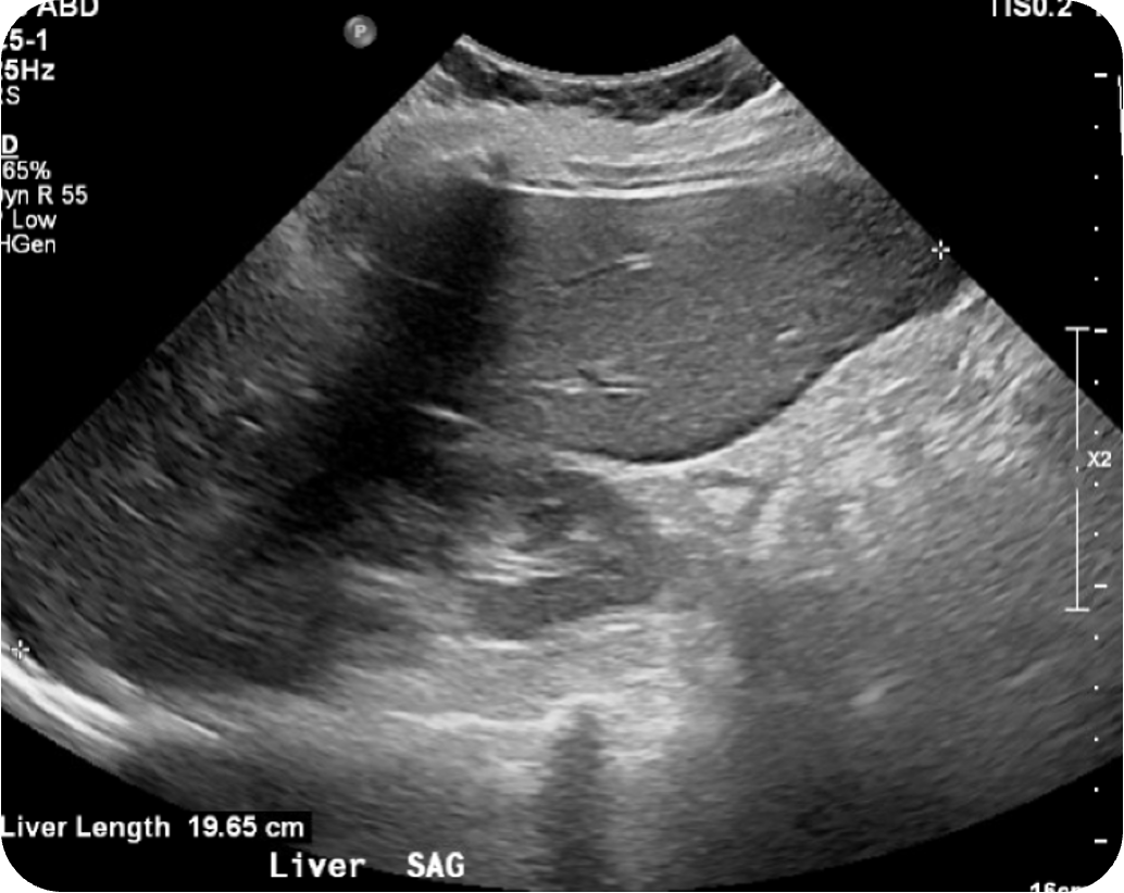
Case Presentation: A 64-year-old woman presented with three months of progressive burning pain and weakness in both calves, leading to wheelchair dependence, accompanied by daily fevers, 10-lb weight loss, anorexia, fatigue, dry cough, and intermittent gross hematuria. Examination showed distal lower-extremity weakness, marked calf tenderness, and mild oral dryness without rash or synovitis. Laboratory evaluation revealed anemia, leukocytosis, thrombocytosis, cholestatic liver injury, and hypoalbuminemia. Autoimmune studies showed ANA 1:640, SSA/SSB positivity, markedly elevated rheumatoid factor, strong MPO-ANCA positivity, and positive smooth muscle antibody, with normal complements and negative cryoglobulins. Infectious work-up was unrevealing. Urine protein–creatinine ratio measured 1.7 g/day with hematuria.Kidney biopsy demonstrated pauci-immune crescentic glomerulonephritis with small-vessel vasculitis and acute tubulointerstitial nephritis. Liver biopsy revealed portal tract arteritis/vasculitis, obliterative portal venopathy, and nodular regenerative hyperplasia, suggesting vascular injury rather than autoimmune hepatitis. The patient was treated with intravenous methylprednisolone followed by high-dose prednisone and rituximab.
Discussion: The clinical picture raised diagnostic uncertainty between primary Sjögren’s syndrome, systemic small-vessel vasculitis, autoimmune hepatitis, infection, and malignancy. ANCA positivity occurs in a minority of patients with Sjögren’s syndrome, but true ANCA-associated vasculitis is rare. Significant proteinuria, hematuria, and biopsy-proven pauci-immune glomerulonephritis supported renal vasculitis rather than classic Sjögren-associated tubulointerstitial nephritis. Hepatic biopsy findings—arteritis, obliterative venopathy, and nodular regenerative hyperplasia—suggested vascular injury patterns consistent with systemic vasculitis or connective tissue disease–associated porto-sinusoidal vascular disease. Together, these findings might indicate a rare Sjögren’s–MPO-ANCA overlap with multiorgan vasculitis.This case emphasizes the hospitalist’s role in recognizing multisystem autoimmune disease, identifying when clinical features deviate from expected phenotypes, and facilitating timely biopsy-driven diagnosis.
Conclusions: This case illustrates several important principles. ANCA positivity alone is not uncommon in Sjögren’s syndrome, but serology cannot establish vasculitis; abnormal urinalysis or elevated urine protein–creatinine ratio warrants early biopsy. Sjögren’s–AAV overlap is rare but increasingly recognized, with MPO-ANCA far more common than PR3. Renal involvement typically manifests as pauci-immune crescentic glomerulonephritis and may be accompanied by neuropathy, interstitial lung disease, or other extraglandular features. Hepatic involvement is atypical and multifactorial; the biopsy pattern of portal tract vasculitis, obliterative portal venopathy, and nodular regenerative hyperplasia reflects systemic vascular injury rather than autoimmune hepatitis. Treatment generally mirrors standard AAV therapy, including glucocorticoids with cyclophosphamide or rituximab, while plasma exchange is reserved for severe pulmonary–renal syndromes. Early recognition of overlapping vasculitis, integration of multisystem data, and timely initiation of immunosuppression are essential to prevent irreversible renal or hepatic damage.