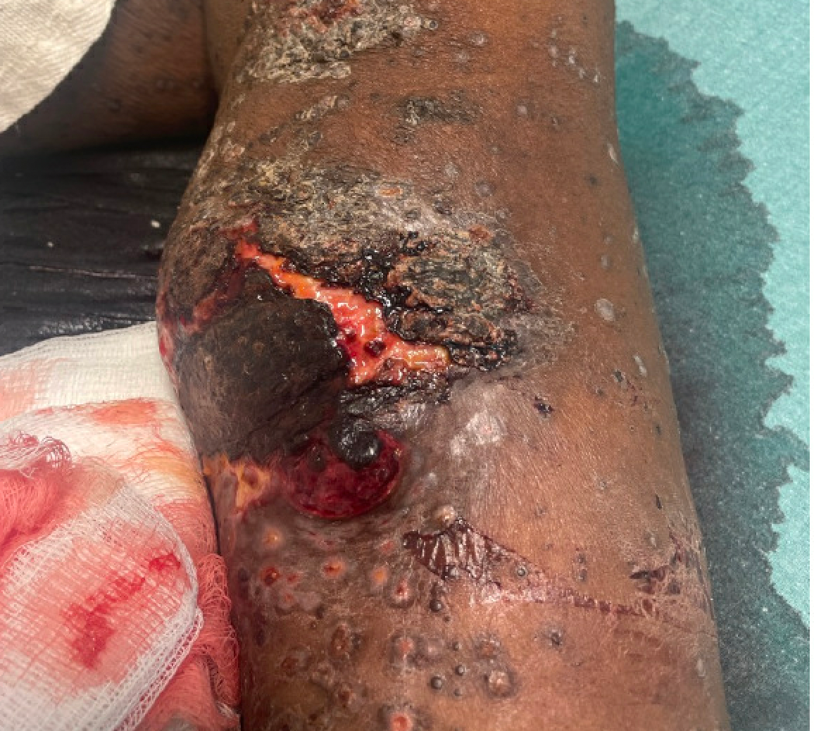
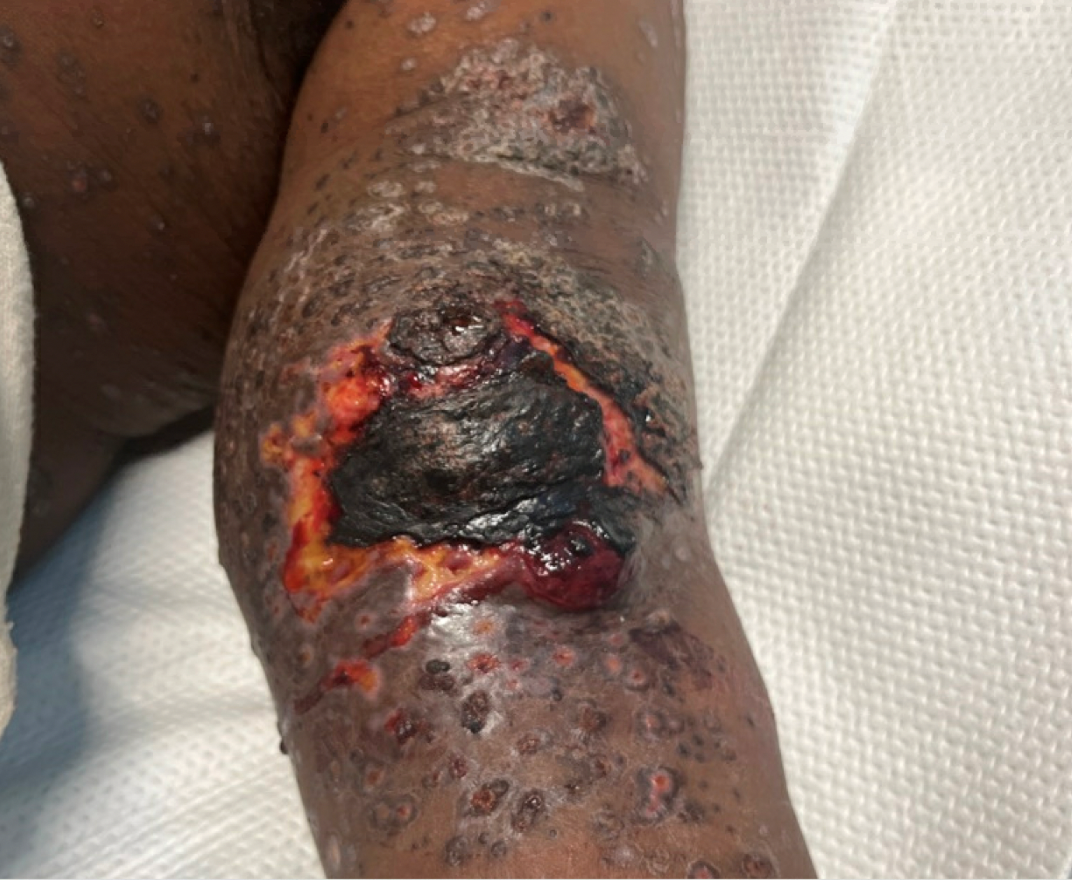
Case Presentation: We present a unique case of a 37-year-old male with a recently diagnosed case of monkeypox three weeks prior and completion of Tecoviramat; who at initial presentation was thought to have an infected arteriovenous fistula (AVF) from a pox lesion likely with an abscess or necrotic process. He had a PMHx of HIV (on ART) and renal transplant (2015) on Mycophenolate and Tacrolimus. His AVF was unused; it had been painful for one month and was bleeding for 3 days, which brought him to the ED. On physical examination, the patient’s left forearm had several coalesced lesions with a large black eschar (9 x 5 cm) with edema and tenderness. There were also generalized lesions along the surface of his body and oral mucosal lesions. He was tachycardic, but hemodynamically stable. Initial laboratory findings included: WBC = 28.76 109/L, BUN = 117 mg/dL, creatinine = 6.7mg/dL, and Hemoglobin = 6.9 g/dL. He was immediately taken for debridement and ligation and empirically started on Vancomycin and Piperacillin/Tazobactam. Surgical findings revealed a large black eschar that extended deep into the venous cuff of the brachial artery. Skin and fatty tissue overlying the AVF had necrosed leaving a black eschar with a thrombus. A residual 24 x 20 cm wound was left open, which was managed by vascular surgery and wound care team. Blood cultures were negative; wound cultures were positive for Methicillin-resistant Staphylococcus aureus and Group G Streptococcus; therefore, antibiotics were continued.
Discussion: Our case portrays an unusual complication of monkeypox causing a cutaneous thrombotic lesion with skin necrosis and erosion into the brachial artery. Some causes of necrotic-appearing skin wounds include gangrene, untreated peripheral vascular disease, hypercoagulable states, poorly controlled diabetes, chronic steroid use, traumatic injuries, and chemical exposures. It is vital to know how to distinguish between a necrotic infected ulcer and a thrombotic lesion to avoid misdiagnosis. Though these lesions have a similar appearance with dark skin discoloration, a thrombosed cutaneous lesion, which may have necrosis due to lack of blood flow may or may not be painful; can present with blackened eschars or dusky, red skin discoloration. Necrotic ulcers have purulent drainage, increased warmth, foul odor along with pain, swelling, and skin discoloration.This patient was urgently taken to surgery out of concern for an infected AVF due to its necrotic appearance. Surgical findings revealed that the necrotic scab was actually a thrombus covering eroded vasculature. This patient’s cutaneous lesion, likely caused by monkeypox, destroyed the surrounding cutaneous vasculature leading to thrombosis and skin necrosis due to disrupted blood flow.
Conclusions: Clinicians should be aware of atypical manifestations of monkeypox in immunocompromised patients, including severe illness, secondary infections, sepsis, hemorrhagic disease, necrotic lesions, and pulmonary involvement. In this particular case, the pox lesions triggered thrombosis of the dermal vessels, causing skin necrosis, swelling and erosion of the brachial AV fistula, causing uncontrollable bleeding. This unique case highlights the importance of understanding the various complications of pox lesions and corresponding prompt management to avoid potentially fatal outcomes.