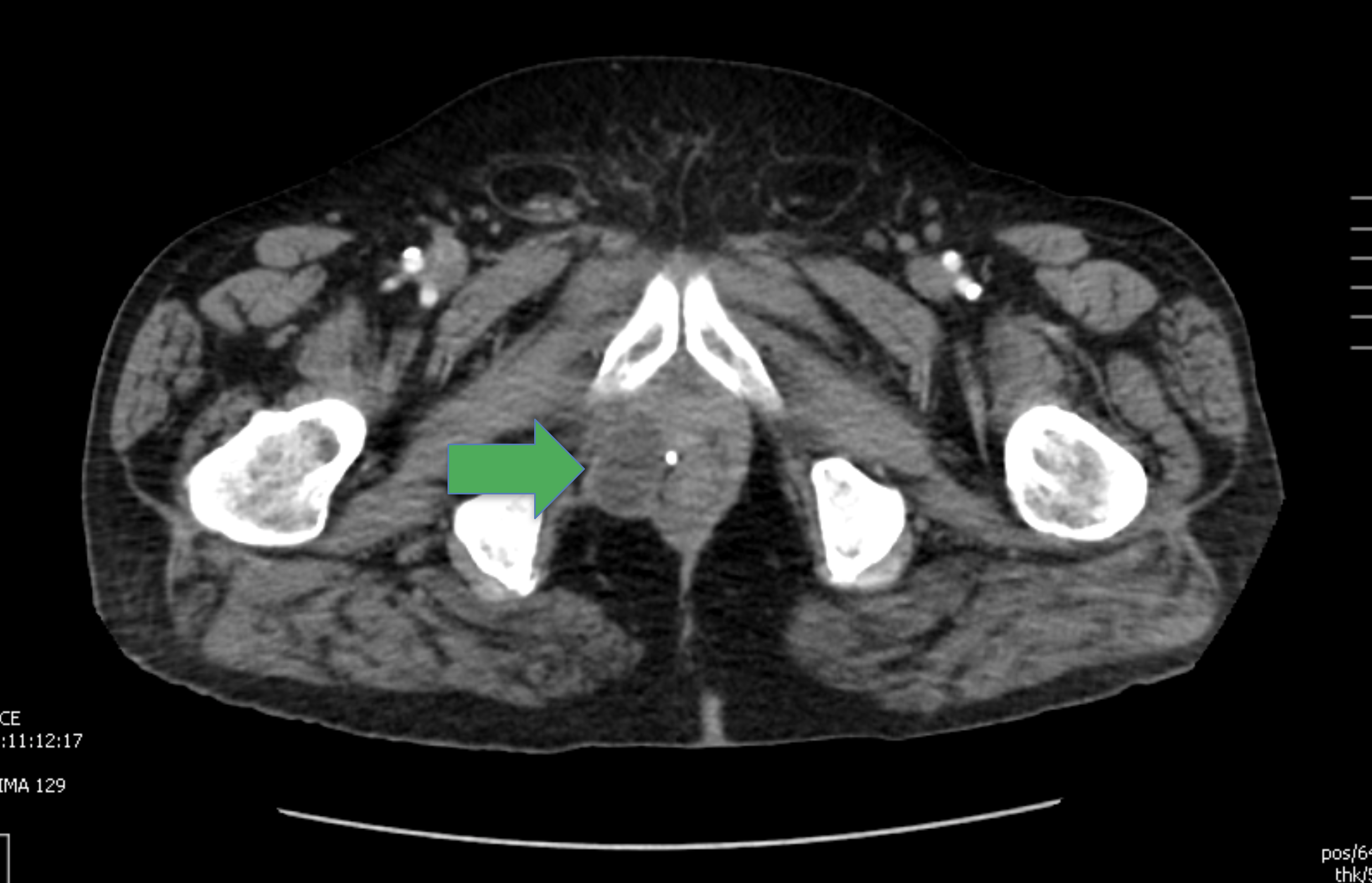
Case Presentation: A 68-year-old male with a past medical history of well controlled diabetes mellitus and chronic lymphocytic leukemia-in remission, presented with a painful right scrotal swelling and redness of four days. The patient denied any previous trauma to the region, urinary symptoms, and recent sexually activity. Ultrasound of the scrotum demonstrated moderate-to-severe epididymo-orchitis. Levofloxacin was initiated but the patient showed no improvement. Due to concerns of a malignancy, a CT scan was done and revealed a hypodense lesion in the right scrotum. Tumor markers including B-HCG, AFP and PSA were negative. A radical right orchiectomy was eventually performed. The histopathology report showed necrotizing granulomas and neutrophilic microabscesses involving the testis and epididymis. Blastomyces Dermatitidis was isolated in the pathological specimen. The patient was started on itraconazole with significant improvement on follow up. On further questioning, the patient denied any travel to any endemic region of blastomycosis.
Discussion: Blastomycosis is one of three major endemic mycoses in the United States along with coccidiomycosis and histoplasmosis. Blastomycosis is caused by Blastomyces Dermatitidis. Endemic regions of blastomycosis in the United States include the Great Lakes, St. Lawrence Seaway, and the Mississippi and Ohio River Valleys. Blastomycosis commonly involves the lungs, but extrapulmonary manifestations can occur, including cutaneous, osseous, genitourinary and CNS infections. Primary genitourinary blastomycosis is very rare and mostly presents with prostatitis. In this case, an immunocompetent host presented with symptomatic primary genitourinary blastomycosis in the form of epididymo-orchitis and without any pulmonary involvement.
Conclusions: Primary genitourinary blastomycosis is very rare, especially that causing epididymo-orchitis. Impaired immunity is a risk factor for extrapulmonary blastomycosis.The broad spectrum of clinical presentations of blastomycosis makes the diagnosis of this condition extremely difficult without a high clinical suspicion.