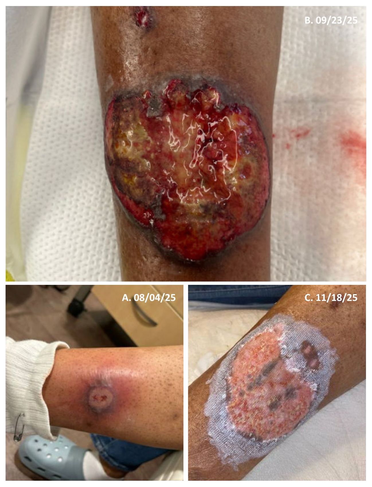
Case Presentation: This is a 65-year-old female with metastatic left invasive breast carcinoma status post left mastectomy and currently receiving chemotherapy, who presented with two months of progressive left lower extremity pain and a non-healing wound. The lesion began as a “pimple” several days after her first cycle of cyclophosphamide, docetaxel, and pegfilgrastim. By her initial evaluation one week later, it had enlarged to approximately the size of a quarter (image 1A). She underwent incision and drainage and was discharged with sulfamethoxazole-trimethoprim; culture grew methicillin-susceptible Staphylococcus aureus. At a subsequent visit, a punch biopsy revealed an ulcerated epidermis with neutrophilic infiltration and tissue necrosis; Gram and Fite stains were negative. Despite therapy, the wound continued to grow, and she developed a new pelvic wound after her second chemotherapy cycle.On admission, examination revealed multiple ulcers on the left lateral leg that encircled a central wound with gunmetal discoloration and raised borders (image 1B). A distinct 1.5-cm, well-demarcated ulcer with an exudative base and violaceous border was present on the mons pubis. Laboratory results were significant for ESR 78 mm/hr and CRP 22.9 mg/L. Pelvic CT demonstrated subcutaneous edema and skin thickening of the left mons pubis, while leg CT showed a 4.6-cm skin defect and nodular cutaneous thickening with inflammatory stranding from the mid-calf to the malleolus. Further workup—including HSV PCR, blood cultures, and MRSA culture—was negative. The presentation was most consistent with drug-induced pyoderma gangrenosum (PG) likely related to pegfilgrastim, supported by characteristic morphology, evidence of pathergy, and clinical improvement with immunosuppression. PG typically presents as rapidly progressive, severely painful ulcers with irregular violaceous borders, peripheral erythema, and a necrotic or purulent base, often beginning as a pustule, papule, or nodule before ulcerating. She was discharged on a prednisone taper, topical clobetasol, dapsone, doxycycline, and wound care after improvement with systemic prednisone. Her wound continued to improve at her follow-up visit with dermatology (image 1C), and her chemotherapy regimen continued without pegfilgrastim.
Discussion: Drug-induced PG is an uncommon subset of neutrophilic dermatoses defined by a temporal relationship to medication exposure. Reported triggers include cocaine, tyrosine kinase inhibitors, isotretinoin, and propylthiouracil. Although colony-stimulating factors such as pegfilgrastim are widely used in non-myeloid malignancies, their association with neutrophilic dermatoses has been described more often with Sweet’s syndrome than PG. In this case, rapid ulcer progression, evidence of pathergy, and a response to immunosuppressive therapy supported the diagnosis of PG. The temporal relationship to pegfilgrastim and lack of alternative triggers further strengthen the association, though causality remains difficult to prove given the rarity of this diagnosis, with only five reported cases.
Conclusions: Pegfilgrastim-associated PG is a rare diagnosis but an important consideration. Clinicians should maintain suspicion for drug-induced neutrophilic dermatoses in patients with characteristic ulcerative lesions after colony-stimulating factor therapy. Early recognition and timely immunosuppressive treatment, paired with appropriate wound care, can lead to favorable outcomes.