Case Presentation: We describe a previously healthy 24-year-old woman who presented with vesiculobullous lesions raising concern for Stevens-Johnson syndrome (SJS) who was subsequently diagnosed with bullous lupus as an initial manifestation of SLE.
24 year old female with PMH of anxiety and depression presented with painful pruritic bullous rash for 2-3 weeks. Blisters appeared first on one of her extremities at the site of a recent tattoo and subsequently spread to her face, upper arms, abdomen and legs. The patient also endorsed fever and malaise. Of note, she presented to different ERs twice, where she was initially diagnosed with possible scabies and eventually with possible SJS and was subsequently transferred to our tertiary care facility for further management.
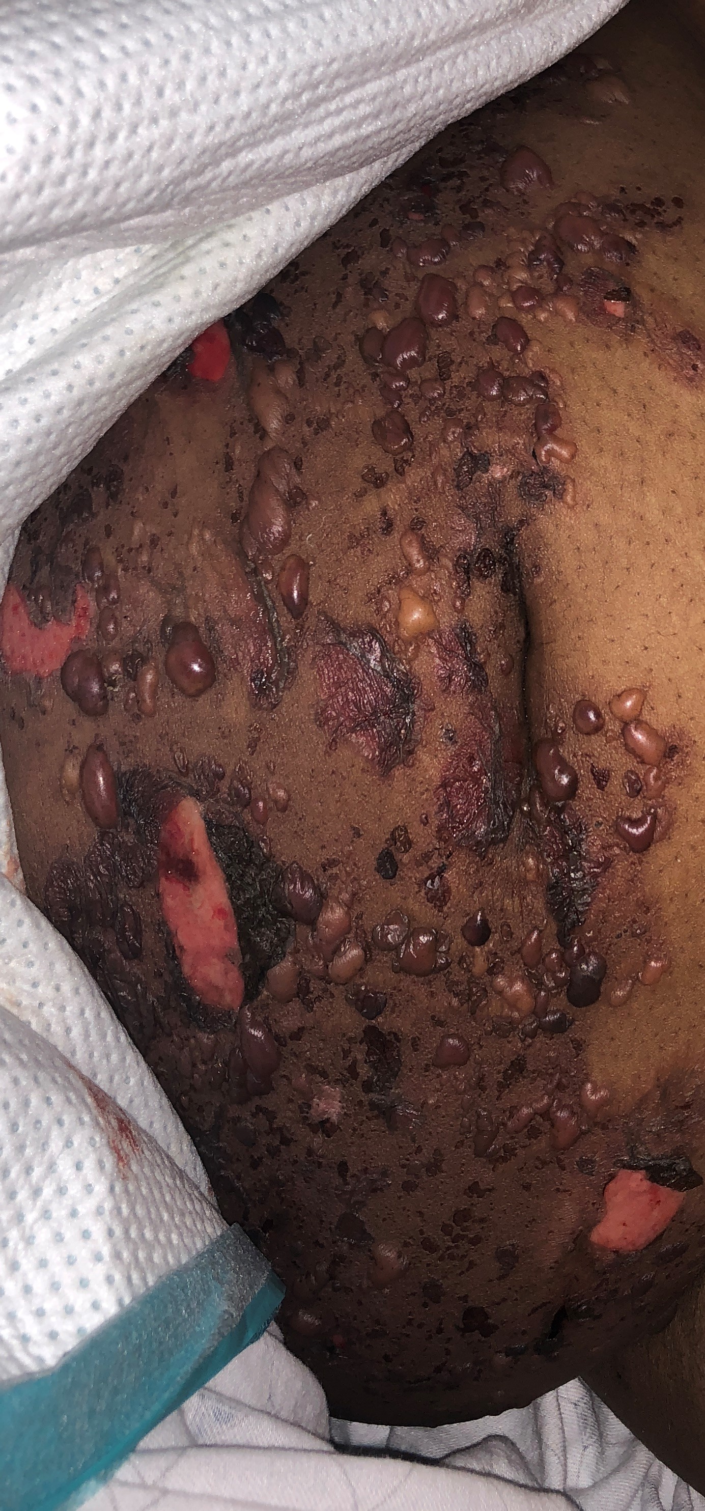
Physical examination showed numerous tense bullae of varying sizes and circular ulcerated lesions with overlying hemorrhagic crust. Numerous oral ulcers and tongue papillitis were detected although no conjunctival or genital mucosal involvement. Symmetric inflammatory polyarthritis was present on joint exam.
Infectious etiology including HSV and VZV were ruled out by negative viral serologies. Drug reaction and SJS-TEN was also considered given prior use of risperidone and involvement of skin and oral mucosa but this was thought less likely because the agent had been discontinued a month prior to onset of symptoms.
Immunologic workup revealed positive ANA, anti-ds DNA, anti-Smith, anti U1RNP and low complement C3.
A skin ‘punch’ biopsy revealed acute cutaneous bullous lesions. Direct immunofluorescence showed strong IgG, C3, IgA and IgM deposition on basement membrane consistent with bullous lupus vs epidermolysis bullosa aquisita (EBA).
The clinical presentation (skin/oral lesions, arthritis and constitutional symptoms), positive SLE serologies and biopsy findings clinched the diagnosis of bullous lupus in the setting of SLE based on fulfillment of SLICC criteria. Treatment with prednisone and dapsone led to dramatic improvement.
Discussion: Bullous lesions are seen in known cases of SLE but only rarely do they occur as the initial manifestation of SLE. This case highlights the importance of awareness of bullous lesions as the first manifestation of SLE. Furthermore, distinguishing BSLE from SJS-TEN is essential as the treatment and prognosis differs substantially. Our patient was repeatedly misdiagnosed which led to significant treatment delay and progression of her symptoms. Dapsone leads to rapid resolution of BSLE lesions with rituximab (B-cell depletion therapy) used as a second-line agent in refractory cases
Conclusions: Bullous lesions may be the first manifestation of SLE and thus it is important to be cognizant of it to come to a timely diagnosis