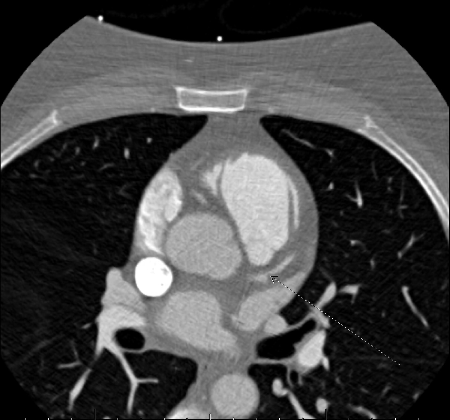
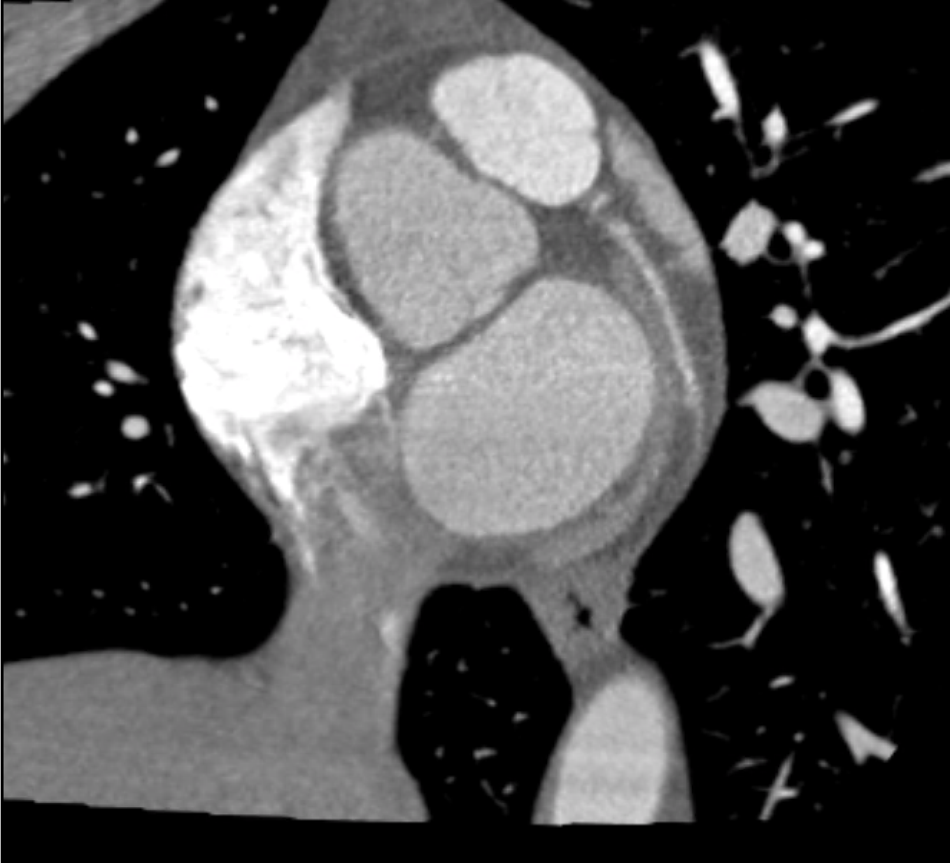
Case Presentation: A 44-year-old obese woman with newly diagnosed hypertension presented with syncope preceded by nausea and diaphoresis shortly after taking her first dose of Nifedipine 60 mg, prescribed two days earlier. She denied chest pain, shortness of breath, palpitations, orthopnea, or edema. On arrival, her heart rate was 140 beats per minute, and ECG showed atrial fibrillation with rapid ventricular response that spontaneously converted to sinus rhythm. Troponin-I was elevated at 45 ng/L, while thyroid and electrolyte levels were normal. Chest X-ray showed no acute findings. Given her new-onset atrial fibrillation and elevated troponin, she was admitted for ischemic evaluation. Transthoracic echocardiogram showed normal systolic function (EF 55–60%) without valvular disease. CT coronary angiography revealed an anomalous right coronary artery (RCA) originating from the proximal ascending aorta, 2.5 cm above the annulus, with an acute-angle takeoff and inter-arterial course showing high-grade dynamic compression between the aorta and pulmonary trunk. No atherosclerotic plaque was identified. Cardiac catheterization confirmed no obstructive disease, though the RCA could not be selectively engaged. Cardiology and cardiothoracic surgery recommended surgical correction via unroofing, translocation, or bypass.
Discussion: An anomalous RCA arising from the left coronary cusp and coursing between the great vessels is a rare congenital anomaly that can mimic acute coronary syndrome by causing myocardial ischemia due to external compression rather than obstructive coronary artery disease. The inter-arterial course predisposes the vessel to compression during systole, leading to transient ischemia and arrhythmias. In this case, administration of Nifedipine, a potent vasodilator, likely reduced coronary perfusion pressure and triggered a myocardial steal phenomenon, exacerbating ischemia in the compressed RCA territory. This case emphasizes the importance of considering structural coronary anomalies in patients presenting with myocardial injury but no evidence of plaque, especially in younger individuals. Standard angiography may fail to capture these anatomic variations, and advanced imaging such as CT coronary angiography plays an important role in diagnosis.
Conclusions: Anomalous RCA with an inter-arterial course represents a rare but potentially life-threatening cause of myocardial ischemia and arrhythmia. Calcium channel blockers may worsen ischemic symptoms in this setting through coronary steal physiology. Early recognition and surgical intervention, such as unroofing or bypass, are essential to prevent recurrent ischemic events and sudden cardiac death.