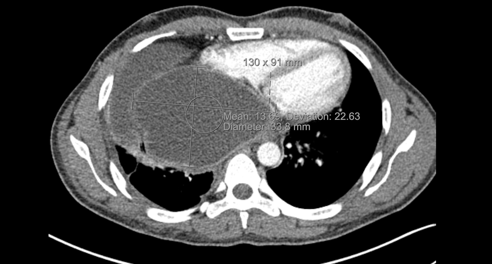
Case Presentation: A 34 year old male presented with abdominal pain and bloating. Over the preceding three months, he developed right upper quadrant pain after eating. His symptoms progressed to diffuse abdominal pain, constipation, and exertional dyspnea. Physical exam showed bilateral 2+ pitting lower extremity edema and 3+ scrotal edema. Labs noted an aspartate aminotransferase 61, albumin 3.0, total bilirubin 2.9. Computed Tomography (CT) chest demonstrated a 13×9x12cm right posterior–inferior mediastinal fluid collection with adjacent rib irregularities. CT abdomen/pelvis showed two cystic collections near the right costophrenic angle without hepatic lesions. TTE revealed a large 8×11cm cystic structure compressing the atria and inferior vena cava. The patient immigrated to the US from Pakistan two years prior to this presentation. Ten years earlier, the patient was told he had developed cysts from ingesting water contaminated with a parasite by sheep and dogs, and hepatobiliary and thoracic surgeries were performed to remove cysts from his liver and lungs. The patient reported no medical therapy after his surgeries. Characteristic imaging findings, immigration from an endemic area, and reported prior cystectomies strongly suggested hydatid disease caused by Echinococcus granulosis. Echinococcus IgG returned positive two weeks after admission. Three weeks after presentation, he underwent right thoracotomy with cystotomy, capitonnage, and cystectomy of a mediastinal hydatid cyst. Albendazole was given 24 hours prior to surgery and postoperative praziquantel was administered to reduce the risk of seeding of new sites. Tissue and fluid cultures were sent for polymerase chain reaction, and the body fluid ova and parasite was negative. Hepatobiliary surgical teams are planning a staged abdominal intervention for the additional right subdiaphragmatic and right abdominal cysts.
Discussion: This case illustrates the importance of integrating historical and clinical assessment in hydatid disease. A notable contributing factor to this patient’s recurrence was the absence of post-operative anti-helminthic therapy following his initial surgeries. Coordinated management involving infectious disease, thoracic and hepatobiliary surgery, and hospital medicine was required to balance diagnostic clarity, symptom severity, and procedural risk. Given that preoperative anti-helminthic management remains inconsistent across guidelines, especially due to a concern that prolonged albendazole use can increase the risk of intra-operative cyst rupture, the team elected to use a short pre-operative course of albendazole (1-3). Though the patient’s cysts exceeded World Health Organization size thresholds for operative intervention, we proceeded with puncture-aspiration-injection-reaspiration strategy in the setting of severe cardiovascular compromise and limited guidance for urgent scenarios (4). Fragmented prior care and the need for multidisciplinary coordination highlight the diagnostic and therapeutic complexity central to hospital medicine (5-8).
Conclusions: Extensive cystic echinococcosis with transdiaphragmatic extension requires individualized, multidisciplinary, patient-centered care that thoughtfully applies existing guidelines while accommodating variability across recommendations. Limited pre-operative albendazole, staged surgery, selective postoperative praziquantel, and prolonged medical therapy may help optimize safety and reduce recurrence.