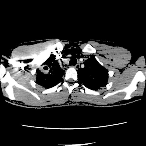
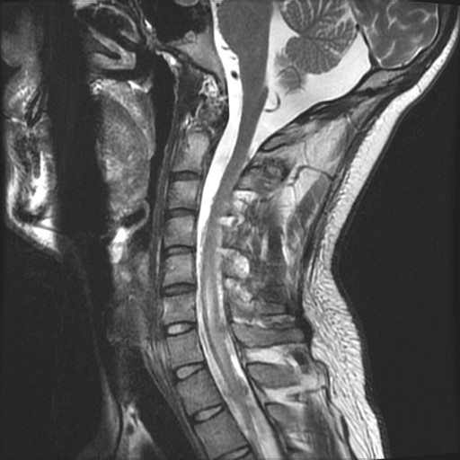
Case Presentation: A 21-year-old male, recently returned from training in the California desert, presented with a non-productive cough and chest pain. Imaging showed a right upper lobe cavitary lesion indicative of pneumonia, and bronchoalveolar lavage was positive for Coccidioides immitis. Treated initially with fluconazole, the cavitary lesion resolved. However, months later, he developed fevers, severe headaches, and photophobia. Meningitis was suspected, but lumbar punctures with PCR and cultures were negative. Blood enzyme immunoassays revealed Coccidioides antibodies, and CSF serology showed complement fixation of 1:16, indicating CNS dissemination.Despite fluconazole therapy, the patient’s headaches worsened, requiring lumbar punctures for elevated intrathecal pressure and VP shunt placement. He was briefly discharged but readmitted with worsening symptoms. Imaging revealed a dural venous thrombus in the superior sagittal sinus. Treatment included apixaban and increased fluconazole (1200 mg daily). Symptoms persisted, with new left arm paresthesia. Imaging revealed progression of meningitis, spinal cord expansion at C6-C7, arachnoid adhesions, and a suspected subarachnoid abscess. Surgical intervention was not required, and treatment escalated to voriconazole and dexamethasone. Voriconazole was discontinued due to transaminase elevation, and posaconazole also caused liver injury. After excluding other causes, IV amphotericin B was started.Amphotericin B was continued until liver function normalized, after which the patient transitioned to lifelong posaconazole (300 mg daily). Symptoms subsided, and he remains under close follow-up.
Discussion: Coccidioidomycosis (cocci), caused by fungi endemic to the southwestern U.S., typically presents with mild respiratory symptoms. In 2017, the CDC reported 14,364 cases in the U.S., primarily in Arizona and California. Males, particularly those with occupational exposure, are more affected, with studies suggesting a 3.5- to 5-fold higher risk of dissemination compared to females. African Americans are also at increased risk of severe disease.Cocci pneumonia is often misdiagnosed as bacterial or viral, delaying treatment and raising dissemination risks. At-risk populations include those with HIV, immunosuppressive therapy, or chemotherapy, though this patient had no such history. This case underscores the importance of considering cocci in patients with neurological symptoms and exposure to endemic regions, even when immunocompetent. CNS involvement can be challenging to diagnose as CSF cultures are often negative. Here, CSF titers, MRI, and epidemiological context were key to diagnosing coccidioidal meningitis. Treatment required escalation to amphotericin B due to liver toxicity from other antifungals.
Conclusions: This rare case of cocci in an immunocompetent male highlights the need for early recognition and multidisciplinary management for optimal outcomes.