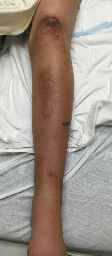
Case Presentation: A 49-year-old woman with a history of D-transposition of the great arteries s/p Mustard procedure, protein losing enteropathy, CVID on IVIG, and chronic anasarca on diuretics presented with redness, swelling, and pain in her left foot. Two weeks prior to admission she fell on a stick and suffered a left shin puncture wound. She removed the stick and noted serosanguinous drainage but did not seek additional care at that time. She had no further issues w/ her leg until 5 days prior to admission. At that time, she noticed erythema and swelling of her left foot as well as sharp pain in the left foot and knee. She treated herself with clindamycin as symptoms were similar to prior cellulitis. Pain worsened and prompted her to present to the emergency department. On presentation she denied fevers, chills, or night sweats. On exam she was afebrile, heart rate 98 beats per minute, respiratory rate 24, and blood pressure 116mmHg/59mmHg. She was noted to be in visible distress and tearful. She had erythema on the dorsum of her left foot and lateral aspect of left thigh with visible puncture wound on anterior shin and exquisite tenderness to palpation and passive movement. Pulses were easily palpable and compartments soft. Laboratory results included a white blood cell count (WBC) of 31,500 per microliter (/uL) with 91% neutrophils, Erythrocyte Sedimentation Rate 53 mm, and C-Reactive Protein (CRP) 8.1 milligrams per deciliter (mg/dL). CT of the left lower extremity revealed diffuse subcutaneous stranding greatest in the medial mid-thigh and posterior medial mid-leg with focal ill-defined fluid but no abscess or subcutaneous gas. She was started on vancomycin, piperacillin / tazobactam, and clindamycin. General surgery was consulted and out of concern for necrotizing fasciitis took patient to the operating room (OR) for incision and drainage. In the OR, there was no evidence of necrotic tissue and compartment pressures were normal. Intra-operative course was complicated by acute-onset, severe pain in bilateral hands, wrists, elbows, knees, and feet for which she was treated with IV hydromorphone. Further lab studies showed a uric acid of 12.1 mg/dL with rising WBC to 48,200/uL and CRP to 25.5 mg/dL. Rheumatology consult team felt that initial symptoms and new polyarthropathy represented a polyarticular gout flare. The patient was started on anakinra with rapid improvement in her joint pain, leg erythema, leg pain, and inflammatory markers. While she completed a course of antibiotics for possible cellulitis, a gouty flare was felt to be the most likely cause of her symptoms and she was discharged on prophylactic febuxostat
Discussion: Hospitalists frequently admit patients with cellulitis that is “refractory” to outpatient antibiotics and must consider a broad differential diagnosis. Gout can mimic cellulitis and cause high WBC with neutrophilic predominance, elevated inflammatory markers, and severe pain. It is important to consider gout as a possible alternative diagnosis, especially if there is concomitant joint pain or other risk factors present, such as the chronic diuretic use seen in this patient.
Conclusions: Though a necrotizing infection is a diagnosis that cannot be missed, in a patient on diuretics who has polyarticular symptoms, severe pain, and signs of severe inflammation; gout should be on the differential diagnosis.