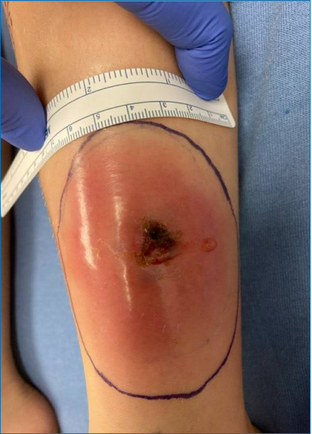
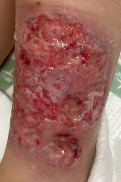
Case Presentation: A 9-year-old female was admitted to a community hospital for intravenous antibiotic treatment of a purulent draining wound on her left shin (Image 1), which had worsened despite completing a 5-day course of oral clindamycin. An ultrasound of the wound showed a 0.8 cm complex hypoechoic collection, prompting urgent incision & drainage for source control and collection of wound cultures. Despite broad-spectrum antibiotics, she spiked a new fever to 101.9°F and her wound continued to worsen (Image 2). Her inflammatory markers continued uptrending, yet her infectious workup remained negative. Upon further chart review and discussion with family, her medical history was notable for a novel auto-inflammatory disease associated with NOD2 mutation, which had previously manifested in infancy as fever, diarrhea, arthritis, and mouth ulcers and was treated with anakinra until the age of 4. Given this history, she was transferred to our quaternary care center for multidisciplinary evaluation by rheumatology, gastroenterology, and dermatology. With signs of multisystem inflammation including rising laboratory markers, anemia, and elevated fecal calprotectin, she was started on methylprednisolone and anakinra with significant improvement in her fever curve, pain, and wound. Esophagogastroduodenoscopy did not show any active lesions, but her colonoscopy showed friable mucosa, loss of vascular pattern, erythema, and erosions in the ascending colon that suggested active inflammatory bowel disease (IBD), for which she received infliximab induction therapy and methotrexate. Having clinically improved, she was discharged 3 weeks after her initial admission with close PCP and specialist follow-up. Since her hospitalization, she has had full resolution of her wounds and continues to follow with gastroenterology and rheumatology for management of monthly infliximab infusions and for monitoring for further autoimmune flares.
Discussion: Pyoderma gangrenosum (PG) is an uncommon non-infectious neutrophilic dermatosis that presents as one or multiple painful ulcers (1). Up to 50% of adults and 74% of children diagnosed with PG are found to have an underlying systemic condition, such as IBD, rheumatoid arthritis, or malignancy; therefore, all patients with suspected PG should undergo a thorough workup that is guided by their medical history and the presence of any other associated symptoms (1,2). Effective treatment of the underlying condition itself is expected to lead to improvement of the PG, as was the case with our patient’s IBD. In cases without a known underlying cause, first-line treatment includes wound care, analgesia, and topical corticosteroids (1). In severe cases, systemic corticosteroids and biologics can be used to achieve faster wound healing, with infliximab currently having the strongest basis of evidence (1). It is important to note that local irritation to the wound can worsen the inflammatory response and prolong wound healing; biopsies and debridement should be deferred unless there is a high enough index of suspicion to investigate an infectious etiology.
Conclusions: A thorough consideration of a patient’s history, presenting symptoms, and laboratory studies can help differentiate pyoderma gangrenosum (PG) from skin and soft tissue infections. The presence of PG warrants further workup for an underlying systemic condition.