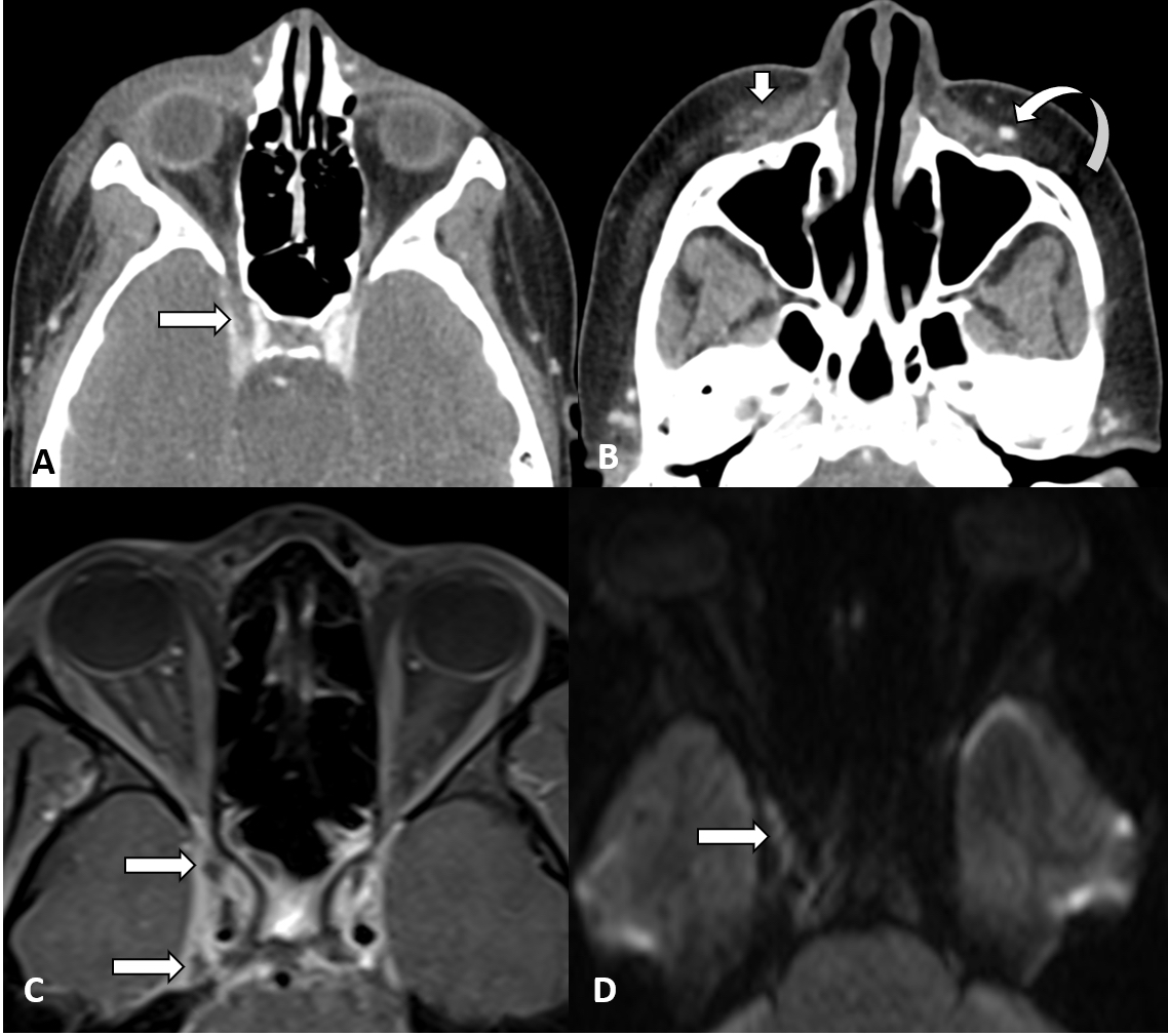
Case Presentation: Our patient is a 15-year-old female, previously healthy, who presented with right facial swelling. She developed swelling of her right upper lip after rupturing a pimple located on her lip. Over the next two days, erythema developed, and the swelling progressed to include her right eye, cheek, both lips, and around her jaw line. She went to her local emergency room (ER) where she appeared acutely ill, therefore was transferred. In our ER, she had diffuse facial erythema and swelling with extension to the surrounding right eye and was febrile, tachycardic, and tachypneic. CT noted preseptal and premaxillary soft tissue swelling with pulmonary nodules in the upper lung consistent with septic emboli. Patient was started on vancomycin and Zosyn. She became hypoxic with new onset chest pain CT-angiogram showed bilateral wedge-shaped defects and septic emboli. At this point, her blood culture was positive for methicillin resistant staph aureus (MRSA). Therefore, she underwent an MRI of the head which showed bilateral cavernous sinus thrombosis. She was treated with vancomycin and enoxaparin. Despite appropriate antibiotics, she remained bacteremic and developed a new pleural effusion, which required drainage. She ultimately was switched to daptomycin to complete an 8 weeks course and enoxaparin shots for 6 months. Patient was doing well on follow-up clinic exams with interval improvement of her septic thrombus on repeat MRIs.
Discussion: This case represents a rare presentation and etiology of bilateral cavernous sinus thrombosis. Extension of these infections is through the veins. Her case is unique because her infection began as acne vulgaris near her upper philtrum. It is commonly believed that spread of the infection is due to the lack of valves in the facial vasculature; however, anatomic studies have shown this is not the case and the primary reason for spread is interconnection of vasculature.Diagnostically, the classic presentation is typically associated with ocular issues such as orbital cellulitis. According to a literature review done by Branson et al, classic septic cavernous sinus thrombosis symptoms of periorbital edema and proptosis occur in 80% to 100% of cases, while headache and cranial nerve palsies occur in 50% to 80%. The only way to confirm the diagnosis is via neuroimaging such as CT or MRI.These infections arise from an initial primary source such as sinusitis, dental abscesses, or facial cellulitis. Thus, identifying the source of infection is critical in determining the organism to treat. The predominant organisms are Staphylococcus aureus and Streptococcal organisms. While cultures are pending, initial antibiotic regiments should be broad-spectrum pending final cultures. Anticoagulation has been controversial in the case of septic cavernous sinus thrombosis as it is rare and the studies have not been definitive. Due to persistent septic thrombosis in our patient, she was treated with treatment dose enoxaparin.
Conclusions: Cavernous sinus thrombosis secondary to infection is a rare phenomenon; however, they can have lethal complications. Early diagnosis with aggressive medical care is the key to decreasing morbidity and mortality. Imaging is a critical component in diagnosis and treatment should focus on antibiotics with some controversy around anticoagulation.