Case Presentation: A 58-year-old Caucasian man with Stage I pancreatic cancer, who underwent Whipple procedure, now presents with 50 pound weight loss over 11 months, abdominal pain, diarrhea, and failure to thrive.
The patient’s symptoms started 6 weeks after the Whipple procedure and progressively worsened. His intermittent nausea and vomiting was occasionally feculent and foul-smelling. Despite increased food intake, caloric supplements, and pancreatic enzyme supplementation, he lost over fifty pounds since diagnosis, with a BMI of 16.5 on admission. His physical exam was remarkable for cachexia with severe bitemporal wasting, angular chelosis, loss of tongue papilla, diffuse muscle wasting, and pallor. His laboratory workup was notable for albumin 1.4 (3.5-5.5 g/dL), prealbumin 6.0 (15-36 mg/dL), potassium 2.9 (3.5-5 mg/dL), and creatinine 0.56 (0.6-1.2 mg/dL). Abdominal imaging (CT, octreotide scan, liver MRI) did not show evidence of malignancy.
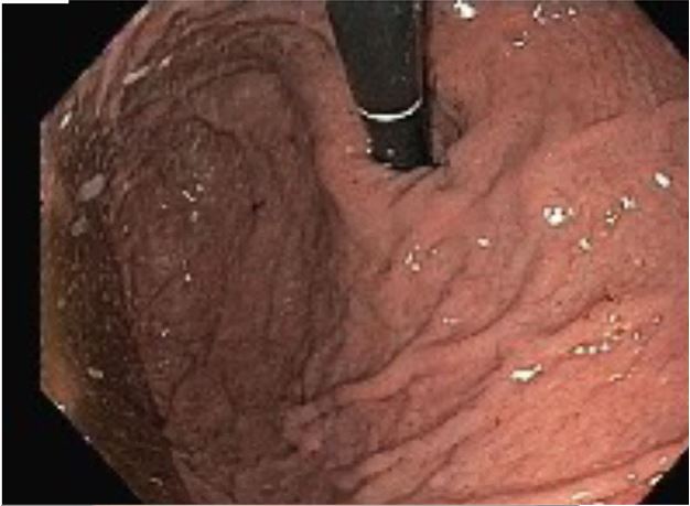
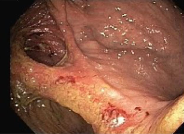
An extensive secondary workup was conducted, including an esophogastroduodenoscopy (EGD) that revealed a deep gastric ulcer, as well as a GCF near the gastrojejunal junction. A nasojejunal tube was placed distal to the fistula to start tube feeds to improve the patient’s nutritional status before surgical correction.
Discussion: Management of pancreatic adenocarcinoma patients, including post-op from Whipple’s surgery, is often transferred to internal medicine physicians. Because these patients’ altered anatomy makes them more likely to have non-specific, recurrent GI symptoms, internists should be prepared to address long-term complications from pancreatic adenocarcinoma, notably those that arise from surgery.
GCF is an uncommon, severe complication of both gastrointestinal (GI) malignancy (occurs <4% of cases) and Whipple’s surgery (3% get peptic ulcer disease (PUD)). The fact that our patient’s fistula likely arose from an ulcer in the setting of pancreatic- not gastric- cancer makes the case even rarer. Our patient’s food was passing from GJ to colon, skipping small bowel absorption and causing the typical GCF symptoms of malnutrition, diarrhea, and feculent emesis. Diagnosis is via barium enema or EGD and treatment involves optimized nutritional status and resection.
The sparse literature on pancreatic cancer-associated GCFs is limited to case reports about post-Whipple ulcers, a finding not present after our patient’s surgery. Furthermore, most post-Whipple patients who developed ulcers did not experience standard peptic ulcer symptoms, making a high clinical suspicion essential in detecting GCF in a pancreatic cancer patient with diarrhea or failure to thrive.
Conclusions: Ultimately, this case presents a unique educational opportunity in terms of identifying PUD and fistula in pancreatic adenocarcinoma patients, and maintaining GCF in the differential for post-Whipple diarrhea and failure to thrive.