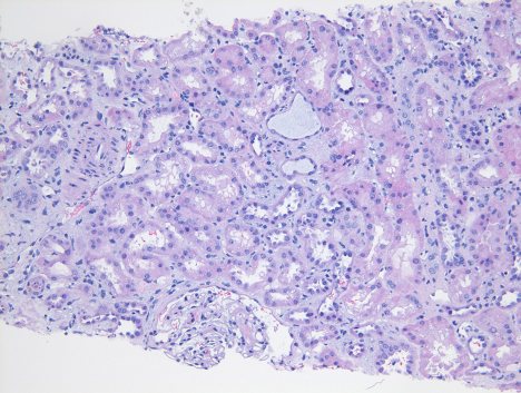
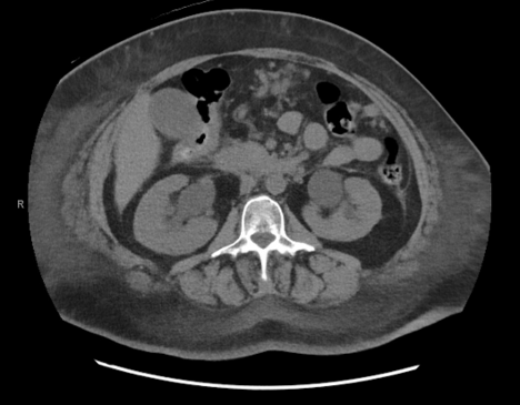
Case Presentation: A 56-year-old female with a history significant for triple-negative breast cancer with peritoneal carcinomatosis, previously complicated by malignant pleural and pericardial effusions, presented to the emergency room with dyspnea and oliguria. She denied preceding trauma or infection, flank or abdominal pain, hematuria, dysuria, and new medications. She had no prior history of kidney disease. Vitals were notable for an oxygen saturation of 88% and tachycardia up to 118 BPM. Physical exam revealed decreased air movement bilaterally on lung auscultation and tenderness in the periumbilical and lower abdominal regions. There was no costovertebral angle tenderness, lower extremity edema, or new skin rash. Initial laboratory studies showed marked serum creatinine elevation (10.0 mg/dL, baseline 0.8 mg/dL, eGFR 4 mL/min/1.73 sq. m) and hyperkalemia (7.1 mmol/L). CT imaging demonstrated stable metastatic disease, mild bilateral hydronephrosis unchanged from CT performed 9 days prior to admission, and persistent bilateral pleural effusions. Patient underwent emergent hemodialysis for the hyperkalemia. Nephrology raised concern for chemotherapy-related intrinsic renal injury, though the oncology team did not think the timeline of her most recent chemotherapy (1 month prior to admission) aligned with renal failure. A kidney biopsy was performed, which did not show evidence of acute interstitial nephritis but was suggestive of acute tubular injury. Concerns remained for obstructive uropathy, but given the lack of significant hydronephrosis, the urology team did not see an indication for immediate surgical intervention. However, the patient remained persistently oliguric despite Foley catheter placement and diuresis, with ongoing rise in serum creatinine post-dialysis. On day 5 of admission, she underwent bilateral ureteral stenting to address ongoing concerns for obstruction from the peritoneal carcinomatosis. Urine output immediately improved with a significant drop in serum creatinine, and the patient was discharged with a creatinine of 1.76 mg/dL.
Discussion: Malignant urinary obstruction is caused by the extrinsic compression or direct infiltration by a malignant tumor into the urinary system, leading to postrenal failure. Diagnosis is challenging in the absence of clear signs of obstruction, such as in cases of non-dilated obstructive uropathy (NDOU). This is a syndrome found in up to 5% of cases of symptomatic urinary obstruction in which there is no or minimal dilation of the pyelocaliceal system on imaging. 74% of NDOU cases have been found to result from malignancy-related causes, including retroperitoneal fibrosis and metastatic disease (1). NDOU has recently been reported in two cases of malignancy-associated obstructive uropathy, with one presumed mechanism of extrinsic ureteral compression or fibrosis causing impaired expansion by the collecting system (2, 3). Diagnostic delay can occur since AKI in cancer patients is often attributed to adverse effects of chemotherapy regimens or tumor lysis syndrome, such as in our patient case (4).
Conclusions: Though it is often assumed that the degree of hydronephrosis correlates with the severity of urinary tract obstruction, it is important to consider the possibility of postrenal etiologies in patients with peritoneal carcinomatosis presenting with a new AKI, even in the absence of significant changes on imaging.