Case Presentation: A 51 year old female with a history of schizophrenia, COPD, and obstructive sleep apnea presented to hospital with complaints of generalized fatigue and acute onset of right-sided extremity weakness. The patient’s family members noted progressive lethargy and confusion on the day of admission. Family members reported the patient’s difficulty with ambulation and increasing right-sided extremity weakness. The patient had complained of headache and falls 1 day prior to presentation but was not able to provide history due to obtundation and confusion.
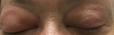
On physical examination, the patient appeared somnolent and withdrawn. Vital signs included a temperature of 102F and HR 94. The patient did not respond to voice commands but did withdraw to pain without posturing. Pupils were pinpoint in size and sluggishly responsive to light. Facial exam revealed bilateral orbital swelling and exophthalmos (image 1). Bilateral ptosis was present. Flaccid paresis of the right upper and lower extremity was present.
White blood cell count was 19.7 x 10⁹/L with 6% band forms. MRI brain imaging revealed acute infarcts involving the left hippocampus, thalamus, midbrain, and pons. MR venogram of the brain revealed cavernous sinus thrombosis extending into the superior ophthalmic veins and left sigmoid sinus thrombosis. Diffuse ethmoid and sphenoid sinus mucosal thickening was seen.
A diagnosis of septic cavernous sinus thrombosis related to sinusitis and complicated by meningitis and acute stroke was suspected. The patient underwent urgent endoscopic sinus surgery with removal of purulent material. An intraoperative microbial culture revealed fungal forms. She was started on broad antimicrobial and anti-fungal therapy as well as intravenous anticoagulation by continuous heparin infusion. She gradually improved and was discharged to a stroke rehab facility after 22 hospital days.
Discussion: Septic cavernous venous sinus thrombosis has a low incidence with around 200 reported cases. The cavernous sinus is a dural venous sinus located laterally to the sella turcica. Infection may reach this space from the face, middle ear, orbit, sinuses and oral cavity. Presenting symptoms typically include headache, fever, and periorbital edema. Cranial nerve findings such as lateral gaze palsy, mydriasis, ptosis, and eye muscle weakness may be present. Exophthalmos may be present. Concurrent ischemic stroke is rare but has been reported and may be attributed to poor venous outflow. Imaging studies include MR venography of the brain which reveals filling defects in the dural sinuses and cephalic veins consistent with thrombosis. Treatment includes anti-microbial therapy aimed at gram-positive, gram-negative and anaerobic organisms, infection source control, and anticoagulation therapy. Anti-fungal therapy may also be considered depending upon culture results. Mortality ranges between 15-30%. In our case acute infectious sinusitis likely led to septic cavernous venous sinus thrombosis, sepsis and acute ischemic stroke.
Conclusions: Due to the catastrophic nature of septic cavernous venous sinus thrombosis, the hospitalist must be alert to the presence of fever, stroke signs and exophthalmos as a presentation of this condition.