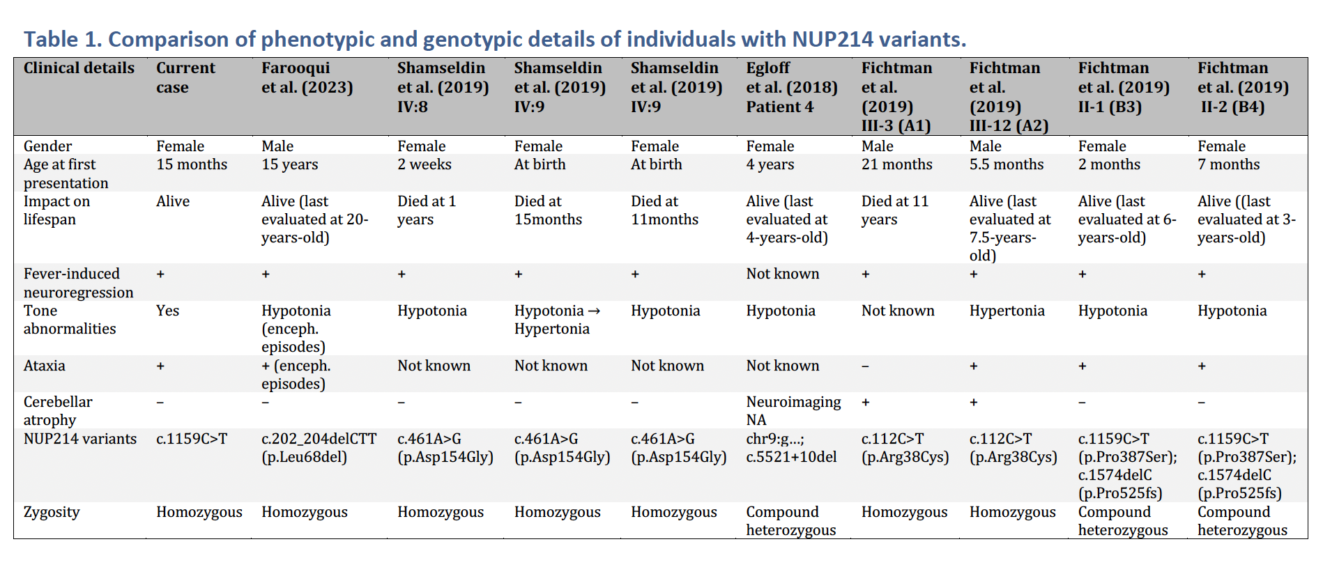
Case Presentation: A 15 month old female with a history of poor weight gain but normal development was transferred for evaluation of atypical seizure-like episodes in the setting of diarrhea and vomiting. Over five days, she had multiple ED visits for abnormal movements—shaking, unusual eye movements, hypotonia and lethargy—some occurring without fever. Initial workup, including head CT, labs, and CSF, was unremarkable. GI panel was positive for Enteropathogenic E. Coli (EPEC). On exam, she had marked ataxia, with inability to sit or coordinate limb movements, and complex eye movements with impaired visual tracking. Video EEG showed no seizures. Brain MRI revealed only a Rathke’s cleft cyst. Ophthalmology and neuroblastoma workups were normal. Despite resolution of GI symptoms, she required NG tube feeds due to persistent hypotonia and poor coordination. Neurology diagnosed acute cerebellar ataxia likely triggered by GI illness, with expected recovery in days to weeks. Given the severity of her regression and the atypical post-infectious inflammatory response to EPEC infection, genetics was consulted. Whole genome sequencing (WGS) revealed a pathogenic variant in the NUP214 gene. At discharge, tone and oral intake had improved, though strength remained significantly reduced from baseline.
Discussion: NUP214 gene variants have been linked to acute encephalopathy and developmental regression following febrile illnesses, typically presenting by early childhood. While symptoms may partially improve between episodes, the condition is often progressive, with early mortality reported. One milder course has been reported, with the patient presenting first in adolescence and showing near symptom resolution between febrile episodes. Clinical features vary but commonly include hypotonia, truncal ataxia, seizures, and abnormal eye movements. Cerebellar atrophy and supratentorial volume loss are common findings on MRI; our patient’s imaging was normal, potentially reflecting an earlier stage of disease.This case illustrates both a rare progressive neurodevelopmental disorder and the utility of WGS in identifying underlying causes of atypical post-infectious complications. While post-infectious syndromes occur, hospitalists have historically lacked tools to explain why some children experience disproportionately severe or prolonged courses or to predict outcomes for prognostic counseling. Recent AAP guidelines now recommend genome sequencing as first-tier testing when the etiology of developmental delay or regression is unclear. Hospitalists should consider WGS or genetics consultation when a patient’s course deviates from expected recovery. In this case, without further investigation, the family may have been discharged with expectations of full recovery, unaware of the potential for persisting deficits or further decline.
Conclusions: This patient’s marked neurological symptoms and developmental regression, in the context of an unusual association between EPEC infection and a post-inflammatory disorder, prompted evaluation for an underlying genetic etiology. Pediatric hospitalists should have a high index of suspicion for genetic conditions when infections are accompanied by atypical, relapsing, or progressive neurological findings. Early diagnosis can guide more targeted management, provide accurate prognostic information, and enable appropriate genetic counseling.