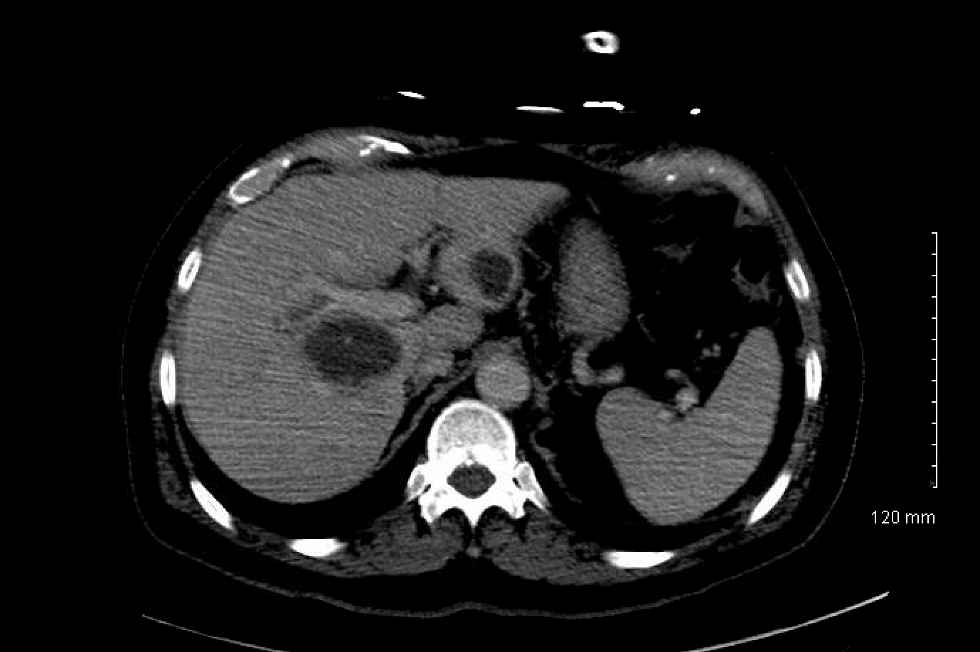
Case Presentation: A 79-year-old man with a past medical history of hypertension and hyperlipemia presented with abdominal pain and fever for two weeks. On physical examination, the temperature was 37.4 C (99.3F), heart rate was 78 beats per minute, respiratory rate was 16 per minute, and blood pressure was 176/81. There was tenderness in the Right Upper Quadrant of the abdomen with no rebounding or guarding. AST and ALT values were, respectively, 66 and 90 U/L. ALP was 138 U/L, bilirubin was 3.9 g/dL, and the white blood cell count was 17.8 K/uL with a neutrophilic predominance. Ultrasound of the abdomen showed small amounts of biliary sludge and cholelithiasis. Murphy’s sign is positive, and findings were concerning for acute cholecystitis. CT scan of the abdomen and pelvis showed multiple fluid collections throughout the liver. Findings are consistent with multiple hepatic abscesses, the largest in the right hepatic lobe’s central/perihilar region, measuring 5.2 x 4.3 x 7.3 cm (Figure 1). An Interventional radiologist placed three percutaneous abscess drain under fluoroscopy guidance, and the fluid culture grew Fusobacterium nucleatum. The patient was treated with intravenous ceftriaxone, 2 g, daily, and oral metronidazole, 500 mg, three times a day. There was no periodontal abscess, and a colonoscopy showed only diverticulosis. Repeat CT done three weeks later showed resolution of the abscess, and the patient became clinically better and was discharged home on metronidazole 500 mg oral TID for two more weeks(Figure 2).
Discussion: Fusobacterium is a Bacteroidaceae family member with 13 species, and Fusobacterium infections have an annual frequency of 0.6-3.5 cases per 1000000 people. Fusobacterium is a non-spore-forming, nonmotile, gram-negative tiny spindle-shaped rod where Fusobacterium necrophorum and Fusobacterium nucleatum are the most prevalent for human illnesses.Fusobacterium nucleatum takes advantage of a FadA adhesion protein to invade and proliferate within host tissues.This cellular entry mechanism disrupts the affected tissues’ permeability, enabling co-infection with other microbes. PLAs can occur as a consequence of colonic pathology as well as a result of periodontal disease. Induction of Fusobacterium nucleatum in mice showed accelerated tumorigenesis through infiltration and upregulation of the NF-B inflammatory pathway. The associated environmental changes increase bacterial growth, leading to increased virulence and tumorigenic effects in the bacterium’s oral and gastrointestinal infections. In our case, the source of the Fusobacterium is cryptogenic, as there was no apparent source of infection. The colonoscopy and the examination of the mouth showed no evident source of infection as there were no periodontal abscesses and colonic pathology, which are the usual sources of the fusobacterium bacteria from where they transmigrate. Image-guided percutaneous drainage is a viable therapeutic option for a single unilocular PLA. Treatment includes abscess drainage as well as antibiotic treatment. Appropriate combination antibiotic therapy consists of a β-lactam and an anaerobic antimicrobial agent (metronidazole, clindamycin). Cultures should preferably be collected before antibiotic therapy, as with other illnesses.
Conclusions: We report a case of Fusobacterium nucleatum presentation with no apparent source of infection, but appropriate surgical drainage of the abscess resulted in symptomatic recovery from the infection.