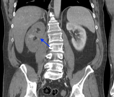
Case Presentation: A 54-year-old male with a medical history significant for hypertension, hyperlipidemia, and type II diabetes initially presented to a local Emergency Department for acute left arm weakness. He had been seen at an urgent care clinic four days prior for upper respiratory infection and prescribed amoxicillin. A non-contrast computerized tomography (CT) scan of the head was unremarkable, and ischemic stroke was diagnosed. However, tissue plasminogen activator was withheld due to platelet (PLT) of 38,000 per microliter. His symptoms progressed to the point of complete left hemiparesis, left hemiplegia, and left facial droop; PLT down-trended to 17,000 per microliter. A CT angiography of the head now showed a thrombus in the right common carotid artery and internal carotid arteries. With no prior heparin exposure, his thrombocytopenia was felt to be due to immune thrombocytopenia (ITP) in the setting of recent amoxicillin use, and dexamethasone was initiated as treatment. The PLT count did not respond to steroids or transfusion, and thus, intravenous immunoglobulin (IVIG) was added for possible steroid refractory ITP. On day three, further work up to evaluate for causes of arterial clots showed low fibrinogen, elevated D-dimer, and prolonged prothrombin time. With these findings, the leading diagnosis became disseminated intravascular coagulation with a concern for underlying malignancy. Subsequently, CT of the chest, abdomen, and pelvis showed no malignancy, but did reveal evidence for a new right renal artery occlusion. Therefore, heparin infusion was started. Later that day, heparin induced thrombocytopenia (HIT) antibody assay that had been sent upon admission returned highly positive at 2.301 optical density leading to immediate discontinuation of heparin and initiation of argatroban. The serotonin releasing assay (SRA) also returned highly positive at 78% in low dose heparin confirming diagnosis of spontaneous HIT. He continued to have thrombocytopenia and was given two doses of IVIG with subsequent normalization of PLT counts by hospital day 10. He was transitioned to warfarin from argatroban before ultimately being discharged to a rehab facility.
Discussion: When patients present with thrombocytopenia and venous or arterial thrombosis clinicians need to have a high clinical suspicion for HIT. Spontaneous or autoimmune HIT in such patients without known heparin exposures has been seen following non-heparin triggers such as orthopedic surgery or respiratory infections. Early consideration of this process should not only prompt the timely and appropriate work-up as HIT labs (anti-platelet factor 4 (PF-4) antibody test and SRA) often have long turn-around times, but should also remind clinicians to avoid heparin products until this work-up has returned. Spontaneous HIT occurs when anti-PF-4 antibodies form in the absence of heparin exposure possibly due to bacterial antigens or due to tissue injury from tourniquet use. Anticoagulation with non-heparin based products has been the standard treatment for all types of HIT, but there is growing evidence for use of IVIG for refractory cases of spontaneous HIT.
Conclusions: This case demonstrates the importance of clinicians having spontaneous HIT on the differential in the setting of unprovoked thrombosis and hemorrhage and avoiding heparin products until spontaneous HIT can be ruled out. In addition to anticoagulation, clinicians should consider IVIG as treatment option for refractory spontaneous HIT.