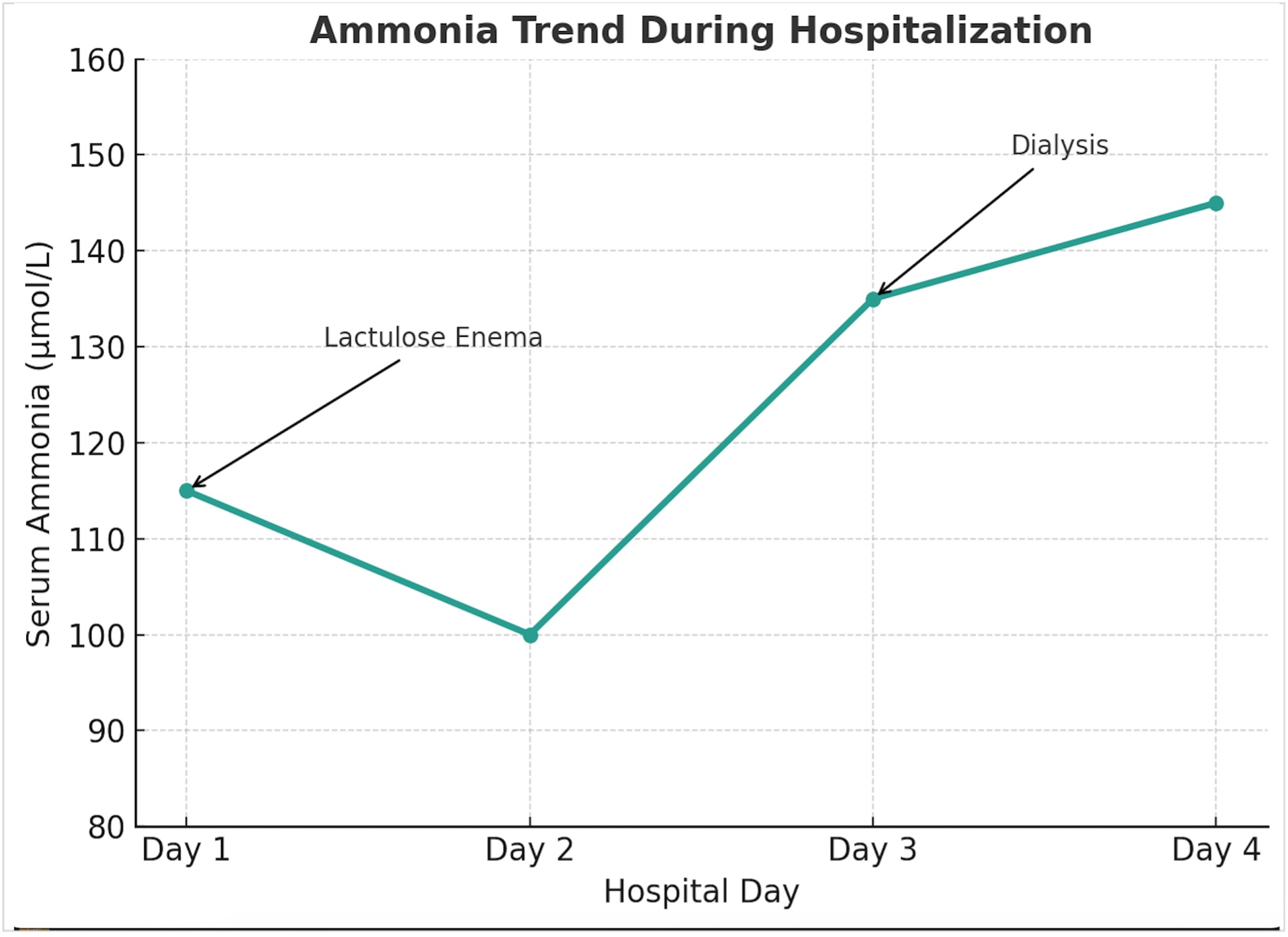
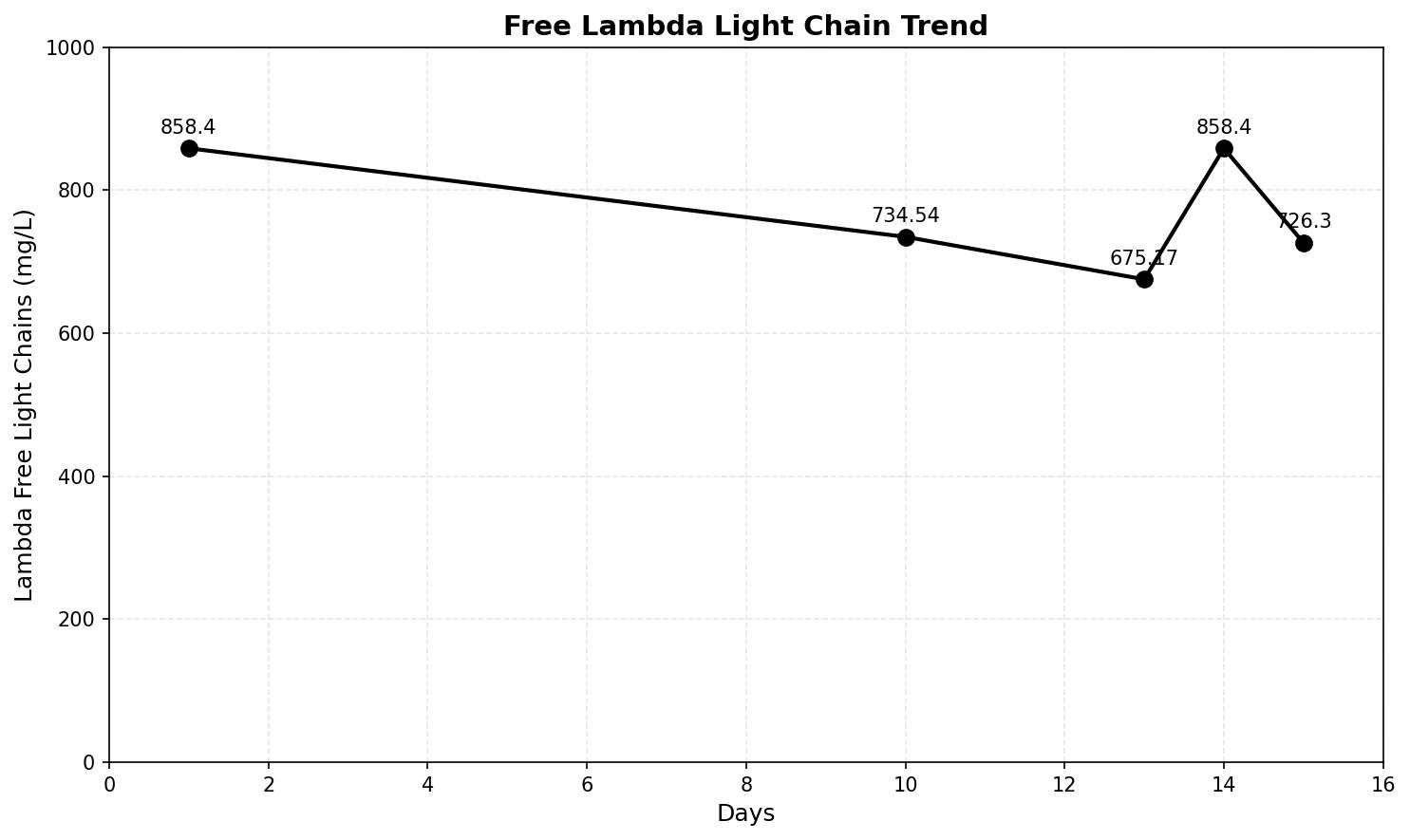
Case Presentation: Our case features a 73-year-old male diagnosed with High-Risk IgG Lambda Multiple Myeloma (MM), initially treated with four cycles of bortezomib-lenalidomide-dexamethasone followed by daratumumab-dexamethasone in anticipation for stem cell transplant. However, his disease was refractory to treatment and continued to progress requiring fourth line treatment with teclistamab. Patient developed altered mental status (AMS) on cycle 2 day 8 of teclistamab, etiology of AMS was initially due to suspicion of grade 2 Immune effector cell-associated neurotoxicity syndrome (G2 ICANS). He was treated with dexamethasone and tocilizumab, with little improvement. Further lab studies showed progression of IgG protein and free lambda light chains, concerning for hyperviscosity syndrome. He started plasmapheresis with some response to IgG and free lambda light chains but without improvement of his AMS. His MRI brain was negative for intracranial pathology and electroencephalograph showed evidence of metabolic encephalopathy without seizures. Additional work up revealed significantly elevated ammonia levels without evidence of hepatic dysfunction, with initial ammonia level of 117 μmol/L. Ultrasound of the liver was negative for hepatocellular pathology. Patient was started on lactulose enemas which had helped decreased ammonia, however rebounded after day 2 of initiating lactulose. Patient was then started on hemodialysis (HD) for hyperammonemia. Although his ammonia fluctuated, it stayed significantly elevated while on treatment without improvement in his mentation. After discussions of goals of care and poor prognosis of refractory hyperammonemia, the family decided to proceed with comfort measures.
Discussion: Hyperammonemia Metabolic Encephalopathy (HME) is not a well-documented complication of MM nor is there a well-defined guideline for treatment of this complication. Though initially considered, G2 ICANS was less likely given the lack of response to steroids. Concurrent suspicion for hyperviscosity syndrome was managed with plasmapheresis, though clinical improvement was inadequate. The patient’s hyperammonemia emerged as the predominant driver of metabolic encephalopathy. Unlike hepatic encephalopathy, it is frequently resistant to standard therapies such as lactulose or HD, and carries a poor prognosis once established. There have been several documented approaches to treatment for patients with HME secondary to MM however most of these did show poor outcomes. In another similar case presentation, cyclophosphamide was given to treat this aggressive paraneoplastic syndrome which helped reduce ammonia to less than 10 μmol/L for five days without any dialysis (3), which supports that antineoplastic agents may be indicated to treat uncontrolled disease. Chemotherapy has been shown to be the only variable associated with improved survival (7).
Conclusions: MM-induced HME should be considered in the differential diagnosis of AMS in patients with advanced MM. Given its low rate of incidence, ammonia is not routinely checked early in the evaluation of encephalopathy as highlighted in other case studies reviewed with similar presentation. This can be an alternate cause of HME in patients with relapsed MM, leading to delay in diagnosis and timely intervention. Increased recognition of this complication and systematic study of management strategies is needed to inform evidence-based guidelines and potentially improve outcomes in this group of patients.