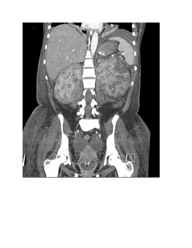
Case Presentation: A 32-year-old woman with history of recurrent urinary tract infections (UTI) presented with malaise and dysuria. Vitals were notable for low grade fever, tachycardia, and hypotension (85/50). Physical exam with 2+ pitting edema and bilateral costovertebral angle tenderness. The white blood cell count was 23,000 cubic mm, creatinine 6.4 mg/dL (baseline 0.6 mg/dL), and ferritin 6,583 ng/mL. Urinalysis with hematuria and normal red blood cell morphology. Blood cultures grew Escherichia coli (E. coli). CT abdomen showed multifocal, bilateral renal abscesses predominantly on the left side. She was started on cefepime and initiated on continuous renal replacement therapy (CRRT). Renal biopsy revealed diffuse macrophage and histiocyte infiltration with scattered calcification, most consistent with malakoplakia. Cefepime was transitioned to IV minocycline, bethanecol and ascorbic acid. She was weaned off CRRT; however, given multiple recurrences of bacteremia without adequate source control, she ultimately required a radical left nephrectomy.
Discussion: Hospitalists commonly care for patients with complicated UTI and pyelonephritis. Though E. coli can cause pyelonephritis, presence of multiple bilateral renal abscesses and renal failure requires hospitalists to look beyond a simple bacterial etiology of pyelonephritis and consider additional underlying processes such as infiltrative, malignant, and inflammatory diseases. Malakoplakia is a rare chronic inflammatory disease that can occur in any organ system though has the highest predilection for the urinary tract system with a 4:1 female to male ratio. Clinical presentation of urinary tract malakoplakia includes recurrent UTI with hematuria and urinary tract obstructive pathology in more advanced disease. The pathogenesis of malakoplakia has three components: presence of infection with E.coli (90% of cases), underlying autoimmune disorder or immunocompromised status such as HIV or transplant status, and macrophage dysfunction in intra-phagosomal digestion leading to granuloma formation. Occurrence of malakoplakia in our patient as an immunocompetent host without known autoimmune disease is rare. Malakoplakia is a histological diagnosis requiring biopsy. Treatment options for malakoplakia vary depending on the site of involvement. In the urinary tract system medical treatment includes fluoroquinolone antibiotics with the addition of sulfonamides or rifampicin. Use of bethanechol with ascorbic acid has also been suggested as cholinergic activity and vitamin C can increase the intracellular cGMP/cAMP ratio in macrophages leading to improvement in intracellular lysosomal function in macrophages. Mass-like growth in malakoplakia can lead to obstructive pathology requiring ureteric stenting, nephrostomy, or radical nephrectomy. In our patient, recurrent bacteremia with higher disease burden of the left kidney necessitated left radical nephrectomy with subsequent long-term suppressive antibiotics while monitoring need for right nephrectomy.
Conclusions: Malakoplakia is a rare chronic inflammatory disease that can mimic inflammatory and malignant processes affecting many organs with predilection for the urinary tract and associated with E. coli infection. Hospitalists’ evaluation of bilateral pyelonephritis with parenchymal destruction and/or infiltration should include consideration of secondary disease processes such as malakoplakia.