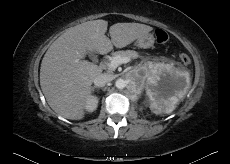
Case Presentation: A 52 year-old woman with a two-month history of metastatic clear cell renal cell carcinoma presented to the emergency department from oncology clinic with one week of malaise and nausea. She also reported subacute onset of dizziness and fever. She received her second cycle of palliative immunotherapy with ipilimumab/nivolumab 14 days prior to presentation. She was uncomfortable-appearing with mild left upper quadrant tenderness but had no abdominal distension, palpable masses, or other focal findings.On presentation, alkaline phosphatase and y-glutamyl transferase were elevated to 315 and 308 respectively, with normal total bilirubin (0.9), aspartate aminotransferase (35) and alanine aminotransferase (31). Hemoglobin was 7.6. Leukocytosis was noted at 11.05 with thrombocytosis to 603. Erythrocyte sedimentation rate was >140. CT scan showed a left renal mass with extension into the renal vein and numerous metastatic lesions in lungs and abdominal lymph nodes. No focal hepatic lesions or intrahepatic biliary ductal dilatation were noted.Initial differential diagnosis in this patient included adverse effects of recent immunotherapy including possible autoimmune sclerosing cholangitis, viral illness, cholecystitis, or paraneoplastic etiology. Given her pattern of laboratory abnormalities, absence of CT-evident hepatic or biliary pathology, and non-specific clinical presentation in the setting of her renal cell carcinoma, a paraneoplastic cholestasis called Stauffer’s syndrome was the most likely etiology of her symptoms. Prochlorperazine and ondansetron controlled her nausea while her dizziness improved with intravenous fluids.
Discussion: Stauffer’s syndrome is a paraneoplastic syndrome with non-specific clinical features often including fever, weight loss, and malaise. Characteristic laboratory findings include elevations in alkaline phosphatase, y-glutamyl transferase, erythrocyte sedimentation rate, and platelets which are not caused by metastasis to the liver. Normocytic anemia is also common. Liver biopsy in these patients often shows cholestasis and possible inflammatory infiltrate. This syndrome is classically seen in renal cell carcinoma but has also been described in prostate, bronchial, and urinary bladder malignancies. These effects are likely mediated by IL-6, a pro-inflammatory cytokine produced by malignant cell lines, as abnormal laboratory values normalize with anti-IL-6 monoclonal antibody treatment. Symptoms and laboratory abnormalities typically persist during active malignancy but improve with nephrectomy.
Conclusions: Hospital medicine physicians frequently see patients with malignancy complaining of non-specific symptoms with many possible etiologies. Stauffer’s syndrome is important to recognize, even in patients without known malignancy. While this was not true of our patient, numerous case reports describe patients presenting with Stauffer’s syndrome who were later found to have renal cell carcinoma. The classic triad of palpable flank mass, abdominal pain, and hematuria is present in less than 10% of renal cell carcinoma patients but up to 20% may have paraneoplastic syndromes. CT scan of individuals with this constellation of symptoms and laboratory values can be crucial in patients without known malignancy, as renal cell carcinoma can be easily missed on abdominal ultrasound often ordered to assess cholestasis. Earlier diagnosis may potentially allow for surgical cure of renal cell carcinoma.