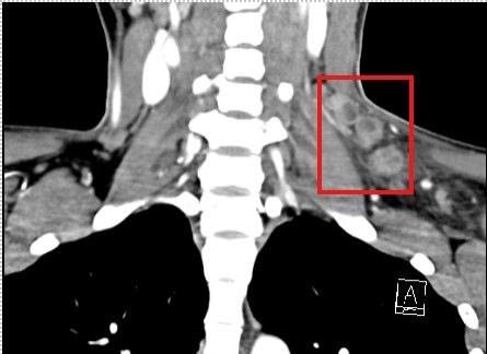
Case Presentation: We present a case of a 22-year-old female with necrotizing lymphadenopathy, later diagnosed with Kikuchi-Fujimoto disease (KFD). The patient initially presented with fevers, night sweats, and painful swelling in the left side of her neck progressively worsening over four days. She also reported lightheadedness, a left-sided orbital headache, and a decreased appetite. There were no preceding symptoms, animal exposures, rashes, recent travel, or known sick contacts. The patient had no personal or family history of cancer.Upon presentation, she was febrile and tachycardic but hemodynamically stable, without signs of respiratory distress. Laboratory results showed a WBC count of 3.4 µg/dL, an ESR of 27 mm/hr, and a CRP level of 35 µg/dL. Physical examination revealed tender, matted supraclavicular and anterior cervical lymphadenopathy. A CT scan of the neck with intravenous contrast demonstrated extensive left-sided cystic and necrotic lymphadenopathy with surrounding inflammatory changes and free fluid (figures 1 and 2). There was no evidence of a primary neoplasm in the head or neck, nor a walled-off abscess. A CT scan of the chest, abdomen, and pelvis showed no significant abnormalities. Due to recurrent severe headaches, an MRI of the brain with and without contrast was performed, revealing non-specific T2 hyperintensity, possibly indicative of an autoimmune or infectious etiology. Empirically, Unasyn and Vancomycin were started. Neurology was consulted, but a lumbar puncture was deferred due to improving neurological symptoms. A broad infectious workup was conducted, yielding negative results for RSV, influenza, COVID-19, tuberculosis, CMV, EBV, HIV, Bartonella, blastomycosis, aspergillus, toxoplasmosis, and syphilis. Autoimmune evaluations were negative for systemic lupus erythematosus, rheumatoid arthritis, and antiphospholipid syndrome. A hematologic workup, including a peripheral blood smear, leukemia/lymphoma flow cytometry, and lymph node core biopsy, was performed. The peripheral smear showed mild leukopenia but no abnormal cell morphology, and the flow cytometry revealed no abnormalities. The core lymph node biopsy demonstrated histopathological features consistent with Kikuchi-Fujimoto disease.
Discussion: Kikuchi-Fujimoto disease (KFD) is a rare, self-limiting condition typically characterized by fever, fatigue, night sweats, painful lymphadenopathy, and, in some cases, skin rashes. The disease predominantly affects young women, especially those of East Asian descent, with the peak incidence occurring in individuals aged 20 to 30 years. The exact pathophysiology of KFD remains unclear, though genetic, viral, and autoimmune factors have been suggested as possible contributors. The diagnosis of KFD is based on distinctive histopathological features observed in lymph node biopsy specimens, which include necrotizing lymphadenitis with scant neutrophils and abundant histiocytes. Treatment for KFD is generally supportive with most cases spontaneously resolving rapidly. Mild symptoms are typically managed with NSAIDs, while more severe or refractory cases may require corticosteroids.
Conclusions: Kikuchi-Fujimoto disease should be considered in the differential diagnosis of lymphadenopathy, especially in young women with characteristic clinical features. A broad differential diagnosis is essential when evaluating lymphadenopathy, with particular emphasis on excluding malignancy, autoimmune conditions, and infectious diseases.