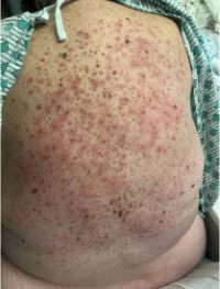
Case Presentation: A 71-year-old woman with a past medical history of hypertension presented for right sided flank pain and post-menopausal bleeding. The initial CT abdomen and pelvis demonstrated bilateral pleural effusions, diffuse lymphadenopathy, and right sided ureteropelvic junction (UPJ) stenosis with hydronephrosis. She underwent lymph node fine needle aspiration (FNA) and then transferred to our institution for urologic and gynecologic evaluations. She arrived with fluid overload and progressive respiratory insufficiency which improved with intravenous diuretics. Echocardiogram demonstrated normal ejection fraction. She completed UPJ stenting with improvement in hydronephrosis. For menorrhagia she underwent transvaginal ultrasound and endometrial biopsy. Perioperative cefdinir was administered. Subsequently she developed a morbilliform rash which dermatology diagnosed as a drug eruption due to cefdinir. However, the novel pruritic rash prompted the discovery of multiple seborrheic keratoses (SKs) spread across her back. Patient stated her skin was clear prior to hospitalization and family confirmed she had no history of SKs. On hospital day 10, the FNA pathology report described poorly differentiated carcinoma with neuroendocrine features, likely gastrointestinal (GI) in origin. This was supported by the endometrial biopsy. She transferred to medical oncology for staging scans and chemotherapy treatment. Her course was complicated by a GI bleed. Scopes revealed discolored, congested mucosa in the gastric body, and a 6-centimeter infiltrative, large mass in the transverse colon. Due to the GI bleeding, and subsequent development of acute deep vein thrombosis, our patient decided to transition to hospice.
Discussion: The Leser-Trelet sign (LTS) refers to spontaneous eruption of multiple SKs that serve as a physical marker of internal malignancy. The sign was first described by Edmund Leser and Ulysse Trelat while studying cherry angiomas in cancer patients during the 1800s (1). LTS is most often associated with adenocarcinoma (67%) and the primary tumor is frequently in the GI tract (72%), followed by breast, lymph system, and lung (4). As in our patient, the physical exam shows numerous SKs, usually in a symmetrical pattern, with the distribution commonly on the back, but the face, neck, abdomen, and extremities may also be involved. Pruritis is the most common complaint (1). The proposed pathophysiology is the production of insulin-like growth factor and epidermal growth factor by malignant cells (3) prompting keratinocyte growth. Treatment of the underlying malignancy results in rapid reduction of growth factor concentration and resolution of at least 50% of the SKs (3,1). This rare paraneoplastic syndrome is controversial because SKs are common in the elderly population and often SK presence is not associated with cancer. However, a few cases describe young patients with LTS whose keratoses resolved with cancer treatment (5). We believe our patient demonstrates LTS since the SKs developed acutely, with associated intense pruritis and GI malignancy.
Conclusions: Although seborrheic keratoses are benign, this case report increases awareness of the potential paraneoplastic process when SKs abruptly develop. Hospitalists encountering this physical exam finding should consider age-appropriate cancer screening given the association with solid organ malignancy.