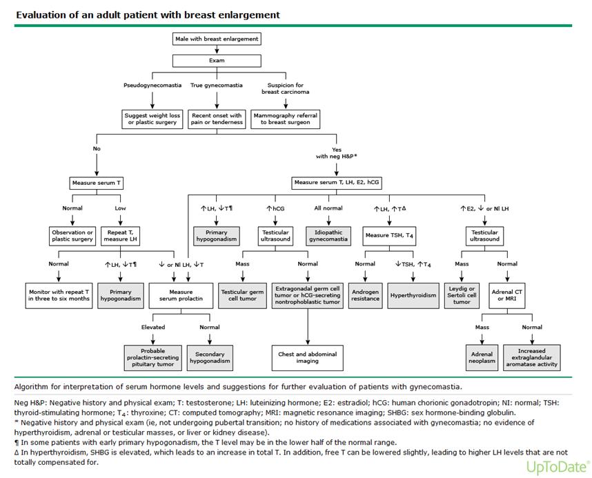
Case Presentation: A 55-year old man with a history of hypertension presented to the hospital with pleuritic left chest pain for 1 day and enlarging bilateral breasts for 2 months. Initial vital signs were notable for elevated blood pressure of 180/90 mmHg. Physical exam was remarkable for bilateral gynecomastia, areolar tenderness and testicular hypotrophy. Admission laboratory results showed normal troponins, elevated prolactin to 45.2 ng/ml, low levels of testosterone 109 ng/dL suggestive of androgen deficiency. Electrocardiogram revealed normal sinus rhythm. Testicular ultrasound did not show any mass and CT scan of the head was unremarkable. MRI of the brain showed a small 0.6 cm non-enhancing right pituitary microadenoma. Patient was started on cabergoline for elevated prolactin and the presence of pituitary microadenoma. Additional laboratory studies revealed normal levels of aldosterone, 17-hydroxyprogesterone, dehydroepiandrosterone; low levels of luteinizing hormone and follicle stimulating hormone. However estrogen level was noted to be markedly elevated at 134 pg/mL. MRI of the abdomen and pelvis was subsequently obtained which revealed a 6.3 cm heterogeneous right adrenal mass extending into the inferior vena cava. The patient’s clinical findings of feminization and adrenal mass with elevated estrogen were concerning for adrenal cortical carcinoma (ACC), specifically feminizing adrenal tumor (FAT). Laparoscopic right adrenalectomy was performed and histological study was consistent with ACC.
Discussion: Functional adrenal adenomas often secrete glucocorticoids, aldosterone, androgen or estrogen. While most adrenal tumors are benign, estrogen-secreting tumors are typically malignant. FAT is exceedingly rare and accounts for only 1-2% of all adrenal adenomas. It can occur either sporadically or as part of hereditary cancer syndromes such as Beckwith-Wiedemann, Li-Fraumeni or MEN syndromes. Male patients often present with gynecomastia, decreased libido, testicular atrophy; female patients present with breast tenderness and dysfunctional uterine bleeding. Diagnosis is based on clinical features and hormonal assessment with marked estrogen elevations. Benign adrenocortical tumors also tend to be <4-cm and appear lipid rich on imaging. FAT have positive aromatase immuno-staining. Treatment includes surgical resection of the tumor, mitotane or aromatase inhibitors and chemotherapy. Prognosis is poor with median survival of 2.9 months after diagnosis.
Conclusions: Male gynecomastia is an uncommon presentation frequently attributed to functional pituitary prolactinomas. However, when sex hormones are elevated, consideration for a concurrent adrenal process is warranted. Clinical judgment is critical in pursuing imaging due to frequently encountered incidentalomas. FAT is an aggressive malignancy with poor prognosis. Early diagnosis and surgical removal are paramount in improving mortality.