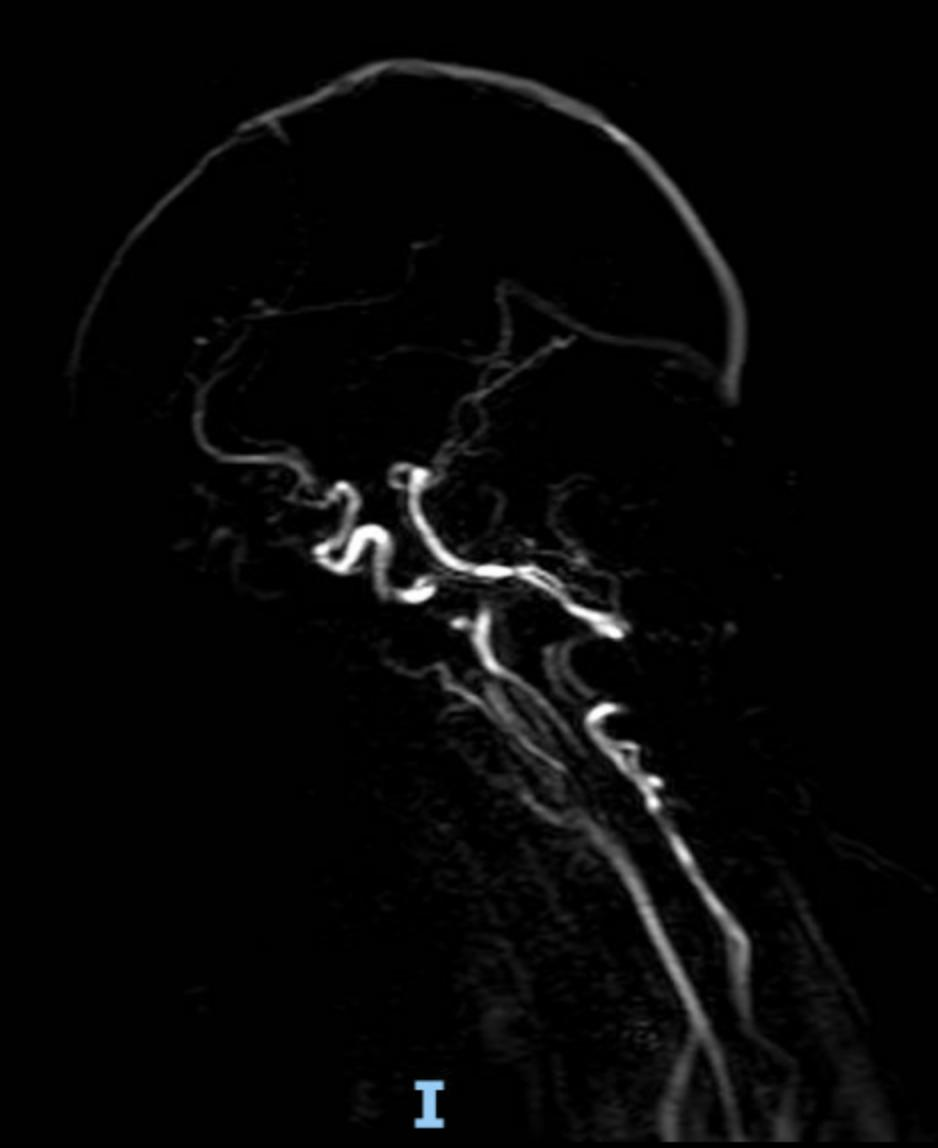
Case Presentation: A 51-year-old female with history of hypertension and systemic lupus erythematous and recent lupus flare presenting with headaches, bilateral hand numbness, and acute onset right sided tremors and weakness of face and hands. The patient also reported some hesitation in her speech which developed overnight. The neurological exam revealed a right facial droop and subtle drift in the right upper extremity with NIHSS of 2. Routine investigation showed normal electrolytes, liver, and kidney function. Cerebral fluid studies also showed 1% lymphocytes, 82 mg/dL glucose, negative CSF paraneoplastic autoantibodies, and negative gram stain and culture. MRI and CT head imaging showed moyamoya angiopathy versus vasculitis. The patient underwent a digital subtraction angiogram showing findings compatible with moyamoya (Fig 1A). There was no vessel wall enhancement to suspect active vasculitis. The patient was treated with aspirin and atorvastatin and continued home medication of prednisone and mycophenolate mofetil. SPECT showing good collaterals and no need for bypass. The patient regained right upper extremity strength and was discharged home.
Discussion: Moyamoya disease (MMD) is a progressive cerebrovascular disorder caused by stenosis of the internal carotid and circle of Willis vessels, leading to formation of fragile collateral networks. This case highlights a rare overlap in which a patient with recent systemic lupus erythematosus (SLE) flare presented with acute neurologic deficits and was ultimately diagnosed with moyamoya angiopathy (MMA). Because SLE itself can cause headaches, stroke-like symptoms, and mood changes, differentiating MMA from lupus vasculitis can be difficult and may delay appropriate treatment.The mechanisms linking SLE and moyamoya remain unclear. Proposed hypotheses include chronic inflammation causing smooth muscle hyperplasia and endothelial proliferation, resulting in luminal narrowing, as well as immune complex deposition or antiphospholipid antibodies promoting thrombotic and proliferative vascular changes. In this patient, CSF studies and absent vessel wall enhancement on angiography made active vasculitis unlikely, preventing unnecessary escalation of immunosuppression.Digital subtraction angiography confirmed moyamoya, and SPECT imaging showed adequate collateral perfusion. She improved clinically with antiplatelet therapy, statin use, and continuation of baseline lupus immunosuppression, without need for surgical bypass. This case underscores the importance of early vascular imaging—particularly MRA, DSA, and perfusion studies—to distinguish MMA from lupus-related vasculitis. A multidisciplinary strategy focused on controlling lupus activity, maintaining cerebral perfusion, and preventing thrombosis is crucial in optimizing outcomes.
Conclusions: This case emphasizes the importance of distinguishing moyamoya angiopathy from lupus-associated vasculitis in patients presenting with acute neurologic deficits. Comprehensive imaging, particularly vessel wall assessment and perfusion studies, plays a pivotal role in guiding management. Early recognition of MMA can prevent unnecessary intensification of immunosuppressive therapy and optimize patient outcomes through targeted, conservative treatment. Multidisciplinary evaluation remains essential, especially in autoimmune patients with atypical cerebrovascular presentations.