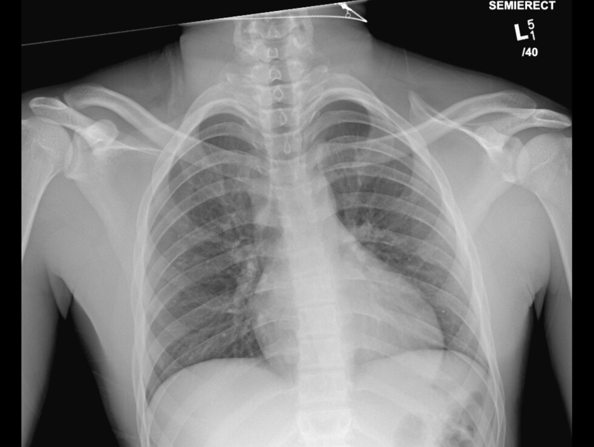
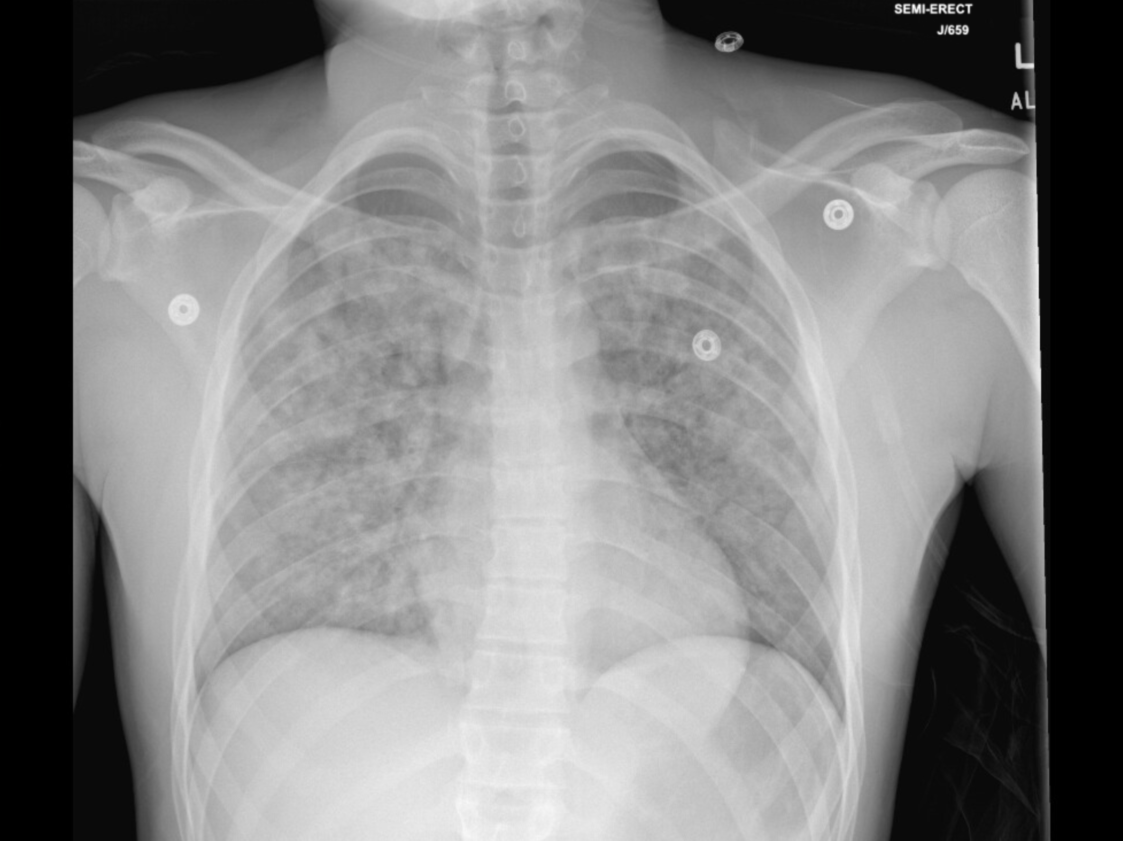
Case Presentation: An 18-year-old male underwent an operation to repair a distal radius fracture. Intubation and surgery were uncomplicated. On emergence from anesthesia, he developed profound hypoxia due to laryngospasm with oxygen saturation dropping below 70%. The laryngospasm was terminated with propofol and patient stabilized in the post anesthesia care unit. However, within hours of returning to the medical-surgical floor, he developed chest pain, tachycardia, hemoptysis, and worsening hypoxia. Repeat chest radiography demonstrated new bilateral diffuse airspace opacities, despite a normal preoperative scan. There was no anatomic airway obstruction on direct visualization, and computed tomography showed diffuse bilateral nodular opacities without vascular injury. Echocardiogram and pro-BNP were normal. The clinical picture raised the concern for negative pressure pulmonary edema (NPPE) complicated by diffuse alveolar hemorrhage (DAH). Management included high-flow nasal cannula oxygen, bronchodilators, intravenous corticosteroids, and diuretic therapy. Positive pressure ventilation was avoided due to risk of worsening hemoptysis. The patient demonstrated rapid improvement with resolution of hemoptysis and hypoxia by hospital day two, allowing oxygen to be weaned and precluded need of invasive diagnostics such as bronchoscopy.
Discussion: NPPE results from vigorous inspiratory effort against a closed airway, significantly increasing negative intrathoracic pressure and pulmonary capillary hydrostatic forces, causing fluid transudation into alveoli. Young, healthy males are particularly susceptible due to high chest wall compliance and the ability to generate extreme negative pressures. While NPPE typically presents with pink frothy sputum, the development of frank hemoptysis and radiographic infiltrates indicates alveolar-capillary uncoupling, consistent with DAH, which is a rare but serious complication. The differential diagnosis for acute postoperative respiratory failure with hemoptysis includes aspiration pneumonitis, pulmonary embolism, acute respiratory distress syndrome, and immune-mediated DAH. The abrupt temporal relationship to laryngospasm, lack of systemic inflammatory features, normal cardiac evaluation, and rapid radiographic and clinical improvement pointed towards NPPE-related DAH as the diagnosis.
Conclusions: This case highlights the complication that is NPPE complicated by DAH, even though it is uncommon, it should be considered in postoperative patients, especially young patients, who develop acute hypoxemia and hemoptysis following airway obstruction. Early recognition, airway stabilization, oxygen supplementation, diuresis, and multidisciplinary monitoring are essential to prevent rapid deterioration. Heightened perioperative awareness of this diagnosis and rapid recognition can facilitate timely diagnosis and more favorable outcomes, as demonstrated by this patient’s rapid two-day recovery. Further research is needed to define optimal adjunctive therapies and long-term implications of NPPE-associated DAH.