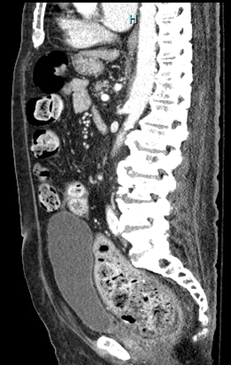
Case Presentation: A 70-year-old male with a history of type 2 diabetes mellitus complicated by neurogenic bladder with indwelling catheter and severe peripheral vascular disease status post bilateral below knee amputations (BKAs) presented with watery diarrhea and nausea for 1 day. Both amputations were in the preceding four months. He required multiple rounds of antibiotics for urinary tract infections (UTIs) during this time period. Physical exam revealed left upper quadrant abdominal tenderness. Labs were largely unremarkable, except for Clostridium difficile PCR being positive. CT abdomen pelvis with contrast revealed a large bolus of stool in the sigmoid colon and rectum, likely representing stercoral colitis, with the remainder of the colon being diffusely filled with stool. Patient was started on a bowel regimen consisting of enemas and suppositories and oral vancomycin for the Clostridium difficile infection, which resulted in clinical improvement.
Discussion: Stercoral colitis is an inflammatory colitis that occurs when chronic constipation and fecal impaction cause colonic distension and the formation of dehydrated masses known as fecalomas. It most commonly affects the sigmoid colon and rectum. The biggest risk factor and most common presentation is chronic constipation. CT abdomen pelvis with contrast is the diagnostic modality of choice. Management typically requires manual disimpaction and an aggressive bowel regimen, but surgery may be required if conservative measures fail. Complications include ulceration, perforation, and ischemic colitis.[1]Clostridium difficile is a gram-positive bacterium most commonly transmitted via the fecal-oral route. The most significant risk factor is antibiotic use, especially clindamycin, cephalosporins, and fluoroquinolones. Typical presentation includes watery diarrhea, fever, nausea, lower abdominal pain, and vomiting. Diagnosis requires detection of Clostridium difficile toxin in the stool. Treatment is determined by the severity of the infection, but often includes oral vancomycin or fidaxomicin. Asymptomatic patients with a positive stool toxin assay do not require treatment.[2]Concurrent stercoral colitis and Clostridium difficile infection is paradoxical and uncommon. The patient above most likely developed constipation due to chronic opioid use in the setting of diabetic neuropathy and decreased gastrointestinal motility, especially given his history of neurogenic bladder, ultimately leading to colonic distension and fecaloma formation. Clostridium difficile infection stemmed from repeated courses of antibiotics for UTIs with a prolonged stay in a healthcare setting, as he required inpatient rehabilitation and skilled nursing following the bilateral BKAs. The diarrhea that the patient presented with was most likely overflow diarrhea from the stercoral colitis rather than from the Clostridium difficile infection, as the colon was noted to be diffusely filled with stool on CT scan.
Conclusions: Stercoral colitis and Clostridium difficile infection occurring simultaneously is rare. A high index of suspicion is required for diagnosis. We report this case to emphasize the importance of not anchoring on a single diagnosis, which would have been easy to do in the above situation, and instead thoroughly investigating and ruling out all potential etiologies.