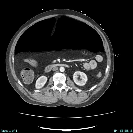
Case Presentation: A 60-year-old male presented after a witnessed syncopal episode. Initial vital signs showed hypertension and tachycardia (BP 143/116 mmHg, HR 132 bpm), tachypnea (RR 30), normal temperature (37.2°C), and hypoxemia (SpO2 84% on room air). Examination showed unresponsiveness despite naloxone administration by EMS, with a tense, distended abdomen. In the emergency department, the patient developed refractory seizure-like activity unresponsive to IV lorazepam, necessitating endotracheal intubation.Post-intubation chest radiograph revealed pneumoperitoneum with subdiaphragmatic free air. CT angiography of the chest, abdomen, and pelvis identified multiple segmental pulmonary emboli, bilateral lower lobe consolidations consistent with pneumonia or aspiration, and large-volume pneumoperitoneum without evidence of visceral perforation.Laboratory studies demonstrated acute kidney injury (creatinine 1.4 mg/dL), hyperkalemia (K+ 5.5 mmol/L), metabolic acidosis (bicarbonate 15 mmol/L), and elevated lactate (4.5 mmol/L), indicating hypoperfusion. Elevated peak airway pressures (39 cm H2O) and hypotension (BP 75/59 mmHg) developed, prompting norepinephrine initiation and urgent surgical consult.Clinical signs of ACS—including tense abdomen, increased ventilatory pressures, and hemodynamic instability—led to emergent bedside abdominal decompression via paracentesis. This intervention resulted in immediate improvement in peak airway pressures (to 15 cm H2O) and blood pressure (to 111/75 mmHg). Exploratory laparotomy revealed no visceral perforation or clear etiology for pneumoperitoneum.The patient’s creatinine peaked at 1.9 mg/dL postoperatively but urine output remained adequate. Extubation occurred on hospital day 8, and discharge on day 12 followed clinical stabilization.
Discussion: Spontaneous pneumoperitoneum, defined as free intraperitoneal air without evidence of hollow viscus perforation or recent abdominal surgery, is a rare but potentially life-threatening condition. It may precipitate abdominal compartment syndrome (ACS), characterized by elevated intra-abdominal pressure leading to compromised organ function. Prompt recognition and multidisciplinary management are essential to avert multiorgan failure and improve outcomes. This case highlights spontaneous pneumoperitoneum as a rare but critical cause of ACS. Conventional causes—such as hollow viscus perforation, trauma, or prior surgery—were excluded. Elevated intra-abdominal pressure impairs respiratory mechanics, venous return, and hemodynamics, hallmark features of ACS. Although intra-abdominal pressure measurement is recommended for diagnosis, clinical urgency limited this. Bedside decompression served as both diagnostic and therapeutic, underscoring the need for timely intervention.Multidisciplinary collaboration between critical care, surgery, and radiology is vital. Literature supports early surgical exploration in spontaneous pneumoperitoneum cases with clinical deterioration, with favorable prognosis if promptly managed.
Conclusions: Spontaneous pneumoperitoneum, while rare, can rapidly progress to abdominal compartment syndrome and multiorgan dysfunction. Early detection, emergent decompression, and coordinated multidisciplinary care are crucial to favorable outcomes.