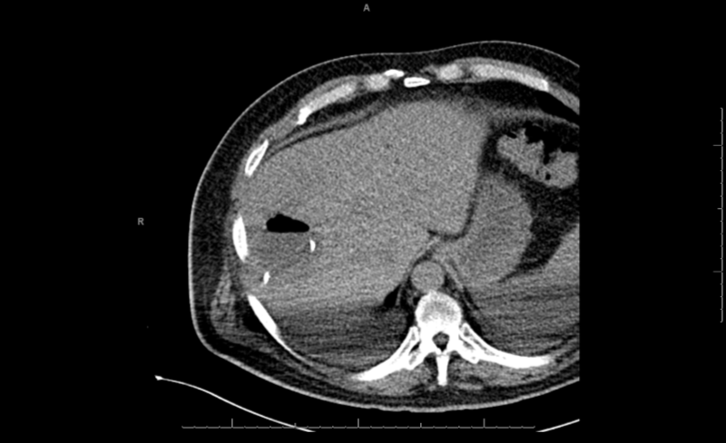
Case Presentation: A 58-year-old male was referred to our institution for outpatient ERCP after suffering an episode of severe hypertriglyceridemia-induced acute pancreatitis complicated by pseudocyst formation at a nearby community hospital five months prior. During that hospital admission, the patient was also diagnosed with type 2 diabetes mellitus. The outpatient ERCP was for a planned Axios stent for drainage of the mature pseudocyst. He had no previous hospitalizations and could not recall ever having antibiotics.The ERCP procedure was notable for stenosis of the ampulla requiring serial attempts before successful cannulation and stenting. He described chills that started the day after the ERCP, followed by right upper quadrant pain, nausea, bilious vomiting, and headache. The patient’s presenting vital signs included a temperature of 38.4 degrees celsius, heart rate of 143 beats per minute, and blood pressure of 131/72 mmHg. The initial physical exam was only significant for right upper quadrant tenderness. His laboratory findings were most notable for hyponatremia, lactic acidosis, hypophosphatemia, and thrombocytopenia. A CT scan of his abdomen showed a 4cm collection of gas within the right hepatic lobe, expected position of the distal common bile duct stent and pancreatic pseudocyst with minimal pneumobilia.The patient was started on piperacillin-tazobactam, intravenous fluids, and admitted to the internal medicine service. Though he initially reported reasonable pain control with acetaminophen alone, the patient acknowledged considerable right upper quadrant pain to the admitting providers and accepted doses of intravenous morphine over the following 48 hours.Although his nausea and vomiting resolved, the patient’s pain, appetite, and laboratory parameters did not improve as expected, as well as worsened thrombocytopenia. On hospital day three, his blood cultures were reported as MDRO E. coli with the OXA-48 class D beta-lactamase gene, a Carbepenem-resistant enterobacteria (CRE) and Clostridium perfringens. He was placed in contact isolation precautions reserved for patients with CRE. His antibiotics were changed to amikacin, ceftazidime-avibactam, and clindamycin. The patient’s abdominal pain, appetite, and thrombocytopenia all improved steadily over the next 72 hours. He was discharged with home intravenous antibiotics to complete a 14-day course.
Discussion: Clostridium perfringens and Carbapenem-resistant enterobacteria are organisms that can quickly lead to septic shock with serious complications, including intravascular hemolysis, renal failure, and death. Mortality rates of bacteremia of 27-44% with C. perfringens [1] and up to 50% from CRE organisms [2] have been reported. Cases of each individual organism have also been reported in the literature following biliary procedures such as laparoscopic cholecystectomy and endoscopic retrograde cholangiopancreatography (ERCP). [3,4]
Conclusions: The incidence of CRE infections has risen worldwide, including outbreaks following ERCP procedures. According to the CDC, there were 9300 cases of CRE (1400 cases of E coli) in the US in 2013.[5] Providers should be aware of the potential for such organisms, especially in the setting of intrahepatic gas on imaging, in patients with abdominal sepsis.