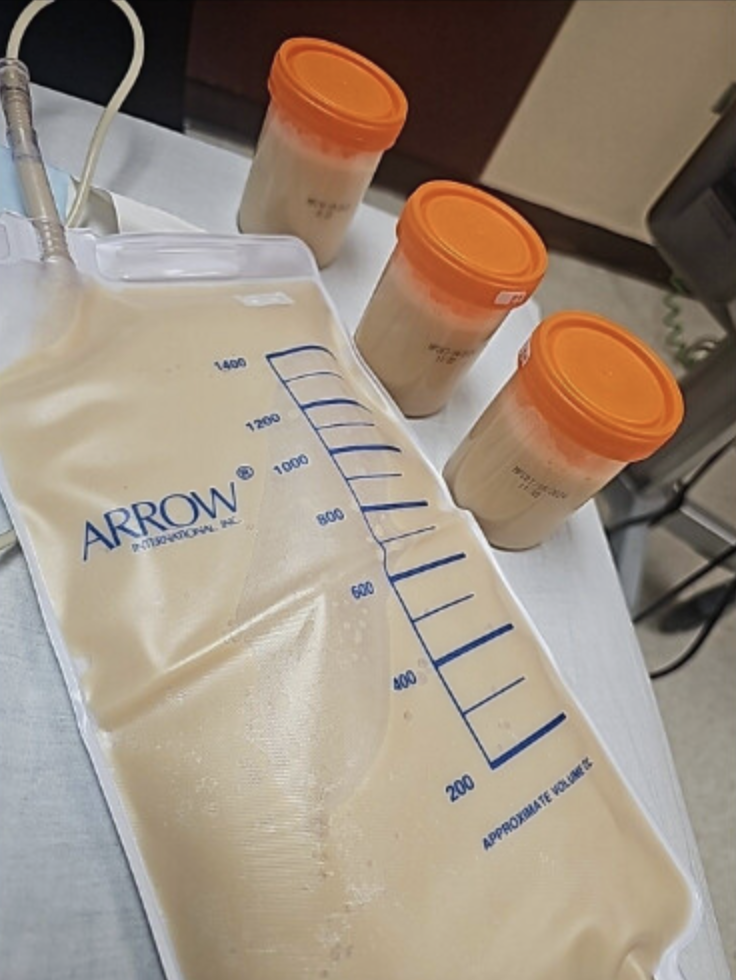
Case Presentation: A 75-year-old female with a history of left radical nephrectomy for renal cell carcinoma presented with one month of progressively worsening left-sided chest and back discomfort following her surgery. CT of the abdomen/pelvis revealed left-sided pleural effusion and a retroperitoneal fluid collection in the left renal fossa. The patient underwent thoracentesis with 600 ml of milky fluid drained. Pleural fluid studies revealed triglycerides of 2182 mg/dl and cholesterol of 151 mg/dl, consistent with chylothorax. Pleural studies were negative for malignancy, and renal mass biopsy had clear margins with no evidence of metastasis, ruling out malignancy as the etiology. Thus, the chylothorax was likely iatrogenic secondary to the left nephrectomy. Percutaneous drains were placed within the left pleural effusion and retroperitoneal collection in the left renal fossa. The retroperitoneal fluid accumulation was also consistent with chylous effusion. The patient was treated with a fat-free diet supplemented with medium-chain fatty acids and octreotide. The output of the drains was monitored throughout the hospital course, and the drains were eventually removed due to minimal output, and no reaccumulation was seen on repeat imaging. Lymphangiography and lymphatic interventions such as embolization were not required, and the patient was discharged on a low-fat diet supplemented with medium-chain fatty acids with pulmonology follow-up to track effusion recurrence.
Discussion: Chylothorax can have non-traumatic causes, with malignancy being the most common, and traumatic causes, with thoracic surgery being the most common. Though less common, thoracic duct injury can also occur in abdominal and retroperitoneal surgeries, as in this case. Translocation of ϲhylе across the diaphragm from retroperitoneal chylе accumulations could be another etiology. Currently, there are no evidence-based guidelines for the management of chylothorax. The etiology of chylothorax, flow rate, and patient condition dictate the preferred management. For patients with low-output ϲhуlοthoraх (< 1L ϲhуle per day), medical therapy with pleural fluid drainage, dietary modification, and initiation of a somatostatin analog may be considered before invasive interventions. Patients with high-output ϲhуlοthoraх (>1L ϲhуle per day), persistent leak, or nutritional or metabolic compromise are typically managed with early operative or invasive therapies.
Conclusions: Chylothorax is an uncommon type of pleural effusion with a broad range of traumatic and atraumatic etiologies. Managing a chylothorax requires a multidisciplinary approach weighing medical therapies and procedural intervention. No management algorithm has been universally adopted for patients with ϲhуlοthorаx since multiple clinical factors impact therapy, including etiology, symptoms, age, functional status, local expertise, and rate of chyle accumulation.