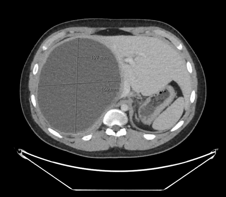
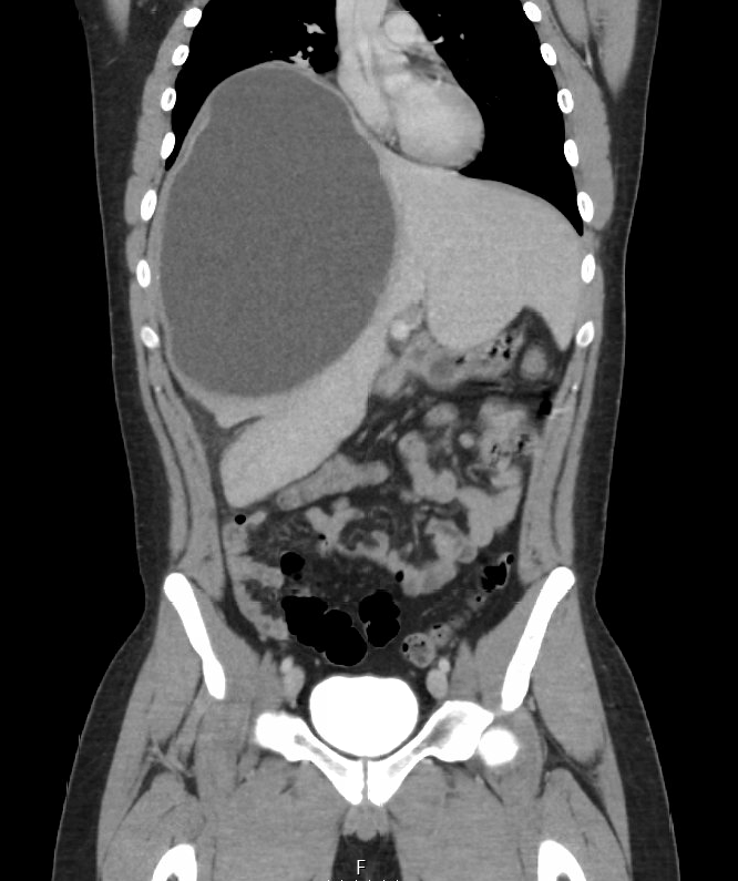
Case Presentation: A 17-year-old male with no significant PMH was admitted with chief complaints of generalized weakness and low-grade fever with recent travel history to Europe. Pertinent labs included leukocytosis 15.9 K and peripheral eosinophilia 1.2%. Chest x-ray showed elevation of right hemidiaphragm. CT showed a large rim-enhancing fluid collection 19.7 x 16.9 x 24 cm in size suggestive of hepatic abscess. He was started on IV antibiotics and underwent CT-guided placement of drain in the right hepatic lobe fluid collection by IR. Repeat scan showed a continuous membrane fold within the collection representing Water Lily sign suggestive of hydatid cyst. Pathology of fluid showed protoscolices fragments of a cestode and Echinococcus serology was positive.There was no evidence of lung involvement. He was started on empiric albendazole and transferred to a higher level of care facility for involvement of a multidisciplinary team. The surgery team deferred resection and he had PAIR procedure done twice with direct infusion of anti scolicidal hypertonic saline. He was discharged on albendazole along with praziquantel for 2 weeks with outpatient follow-up with ID with serial abdominal ultrasounds every 3 months and yearly CT lung monitoring. Despite completing 6 months of medical therapy he continued to have intermittent episodes of fever without evidence of any other infectious etiology. MRI of the abdomen redemonstrated a large 18 cm hepatic cyst. He is currently on life long treatment with albendazole and is undergoing evaluation by hepatobiliary surgery for possible surgical intervention.
Discussion: Echinococcosis is a parasitic infection caused by tapeworms and affects 2-3 million people globally. Major clinical presentations include cystic, alveolar and polycystic echinococcosis. Cystic Echinococcosis is caused by Echinococcus granulosis and mostly involves liver and lung. Infection is acquired by ingestion of eggs through handling of dogs or contaminated food or water. Disease is mostly prevalent in North East Africa, South America and cases in Western countries are rare. Patients are usually asymptomatic for years given the slow growth of cysts. Symptoms vary depending upon the number, size and location of the cysts. Cyst rupture may present with fever, hives or anaphylactic shock. The mainstay of diagnosis is imaging studies including ultrasound, CT or MRI. Lab work can provide supportive data including serology and eosinophilia. Therapeutic interventions include watchful waiting versus medical therapy and PAIR procedure(puncture, aspiration, injection, reaspiration). Laparoscopic or open surgery might be needed for large or complicated cysts. WHO ultrasound classification provides guidance for determining the preferred intervention. Patients require long term radiologic follow up as relapses frequently occur. WHO guidelines for treatment of patients with cystic Echinococcus recommend addition of praziquantel in patients when cystic content spillage is suspected to increase protoscolicidal activity of albendazole.
Conclusions: Physicians should have a high index of clinical suspicion in patients from endemic areas for early diagnosis. Patients require multidisciplinary management involving surgery, interventional radiology and infectious disease to combine both medical and surgical therapies. It is especially important to remember that cyst rupture can lead to anaphylactic shock with fatal outcomes and any surgical intervention should be cautiously coordinated.