
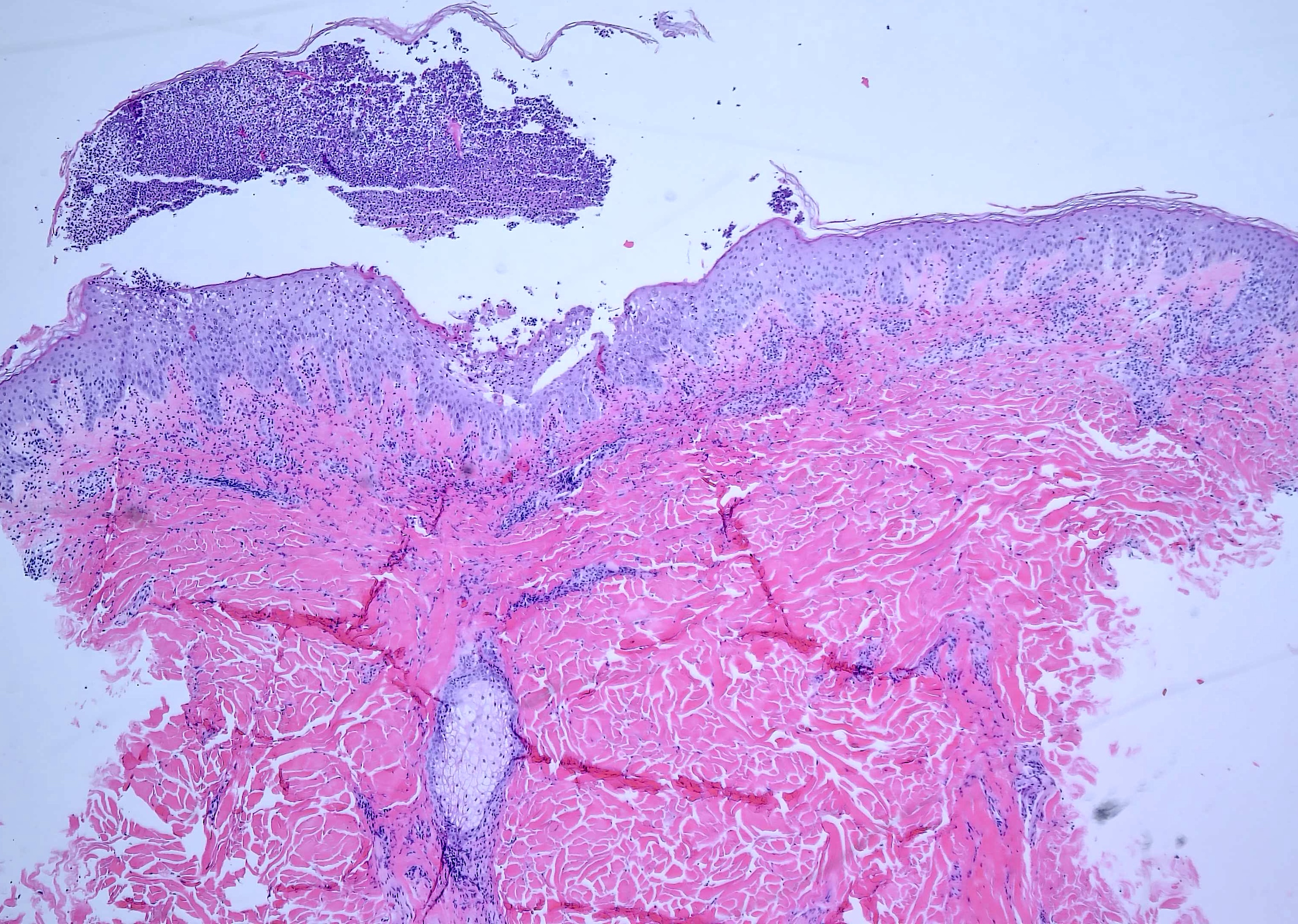
Case Presentation: A 55-year-old woman with diabetes, hypertension, and hyperlipidemia presented with a rapidly progressive pustular eruption of one week’s duration. Two months earlier, she had been treated with terbinafine and doxycycline for presumed onychomycosis, during which nail dystrophy, dactylitis, and bullous lesions were noted. Four weeks into therapy, she developed urticarial plaques and pustules on the trunk. Skin biopsy revealed subcorneal pustular dermatitis, leading to terbinafine discontinuation for presumed acute generalized exanthematous pustulosis (AGEP). Despite medication withdrawal and topical corticosteroids, the rash worsened.Within five days, she presented to the hospital with confluent erythematous plaques and pustules involving the trunk, extremities, and face, with desquamation and severe pruritus but no fever or mucosal involvement. Broad-spectrum antibiotics and systemic corticosteroids were initiated without improvement. Following steroid taper, her eruption worsened dramatically. Repeat biopsy showed psoriasiform epidermal hyperplasia, parakeratosis with neutrophils, and loss of the granular layer, findings consistent with pustular psoriasis. The presence of nail dystrophy, dactylitis, and steroid-related worsening supported a diagnosis of generalized pustular psoriasis (GPP). Corticosteroids were discontinued, and cyclosporine (3 mg/kg/day) was initiated, resulting in marked improvement within days. She was considered for intravenous Spesolimab, an IL-36 receptor antagonist recently approved for GPP, but due to cost barriers, treatment was transitioned to Risankizumab (IL-23 inhibitor) with complete resolution of the rash.
Discussion: This case underscores the diagnostic and therapeutic complexity of acute pustular eruptions frequently encountered in hospitalized patients. AGEP and GPP share overlapping clinical and histopathologic features, yet management differs drastically. AGEP is a self-limited drug eruption that resolves with cessation of the offending agent, whereas GPP is an autoinflammatory psoriasis variant requiring systemic immunosuppression. Misclassification can lead to inappropriate corticosteroid use, which may exacerbate GPP and precipitate life-threatening flares.Our patient represents a clinically challenging and instructive case, not only due to the abrupt and fulminant presentation but also because terbinafine is an exceptionally rare trigger of GPP. Awareness of such uncommon associations is essential, as delayed or incorrect treatment can lead to morbidity, prolonged hospitalization, and systemic complications. The rapid response following cyclosporine initiation underscores its value as a fast-acting immunosuppressant that targets T-cell activation and cytokine release. Limited access to Spesolimab, the first FDA-approved GPP therapy, positions IL-23 blockade with risankizumab as a potential alternative.
Conclusions: Generalized pustular psoriasis can closely mimic acute drug eruptions such as AGEP, leading to diagnostic uncertainty and inappropriate therapy. In hospitalized patients with pustular eruptions, clinicians should maintain suspicion for GPP, especially when lesions persist after drug withdrawal or worsen with corticosteroids. Early dermatology consultation, histopathologic confirmation, and prompt initiation of systemic immunosuppressive therapy can be lifesaving.
.png)