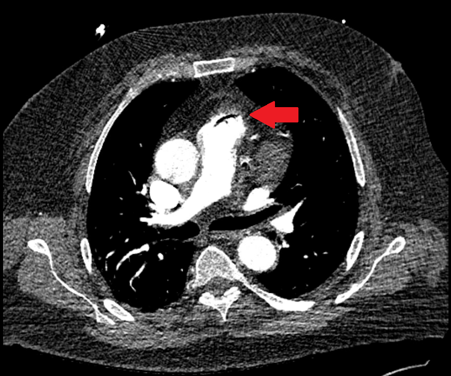
Case Presentation: A 69-year-old male with history of iatrogenic pneumomediastinum after EGD (2 months prior) presented to the ED with 3 days of watery nonbloody diarrhea, vomiting, and fever up to 104°F. His Apple Watch alerted that his heart rate was 120 and O2 saturation 80%. He denied SOB or chest pain. In the ED, initial SpO2 89% on RA, 93% on 2 L, HR 120, BP 121/78. WBC 6.1 (36% bands), Hgb 17.3, creatinine 2.0, lactate 1.9. CTA chest revealed a small amount of gas within the anterior proximal main pulmonary artery compatible with a gas embolism. [Fig 1]He was admitted to Hospital Medicine. CT abdomen/pelvis showed acute colitis involving the ascending and transverse colon but no pneumatosis or portal venous gas. Echocardiogram was normal. Air embolism was managed with supplemental O2 via high flow nasal cannula. He was placed in left lateral decubitus (LLD) position and given IV fluids. By hospital day 2, he was weaned off O2. Colitis was treated with ceftriaxone and metronidazole. Stool GI pathogen panel was positive for Campylobacter. Antibiotics changed to azithromycin. Diarrhea and fevers resolved. He was discharged home on hospital day 4. Two months earlier, he had outpatient EGD and colonoscopy. A duodenal polyp was resected. Post-procedure he acutely developed subcutaneous emphysema at the left side of his face, neck, and chest. CT showed pneumomediastinum extending into the neck and moderate left sided pneumothorax. No CT evidence of bowel perforation. He was admitted for 3 days and managed conservatively. Barium swallow was negative for contrast leak. The subcutaneous emphysema, pneumomediastinum, and pneumothorax resolved. After discharge he was asymptomatic until the present admission.
Discussion: This patient presented with an unusual case of venous air embolism in the setting of Campylobacter infection, with history of an EGD complication two months earlier. Pneumomediastinum and subcutaneous emphysema is a rare but reported complication of EGD.[1] Duodenal polyp resection or mucosal injury by the endoscopic tip may have allowed entry of insufflated air into the luminal wall. He recovered from the initial hospitalization, but enteral inflammation caused by Campylobacter may have allowed venous entry of air via an existing mucosal defect. However, CT abdomen/pelvis did not show free air or evidence of perforation.Air embolism is a rare complication of endoscopic procedures, but usually presents as a sudden hemodynamic instability during endoscopy.[2] In this case, air embolism occurred two months afterward. This patient did not have another source of air embolism, such as other surgeries, IV catheters, chest trauma, or barotrauma.[3]Hospitalists should recognize that air embolism is mainly treated with supplemental O2 with high FiO2. The higher partial pressure of O2 reduces the partial pressure of nitrogen in the blood, which causes nitrogen to diffuse out of the air bubble, reducing its size.[3] LLD positioning allows air to move superiorly into the right ventricle. IV fluids reduce further entry of gas into the venous system by increasing venous pressure. Mechanical ventilation and/or hyperbaric O2 may be needed for unstable patients. This patient’s air embolism resolved with high flow O2 and LLD positioning alone.
Conclusions: This patient presented with venous air embolism during an acute diarrheal illness. Prior mucosal injury from EGD have been reaggravated by the Campylobacter infection, resulting in the air embolism. He fully recovered with high flow O2 and antibiotics.